How a Cardiac CT Scan Helps Detect Coronary Artery Disease
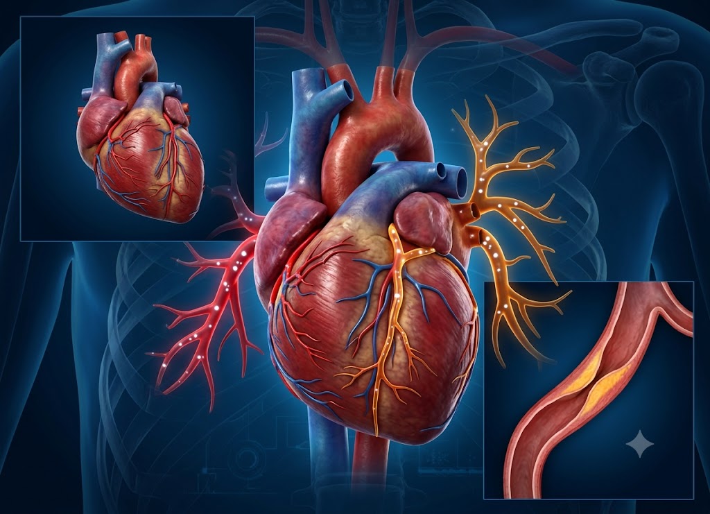
A cardiac CT scan is a painless imaging test that combines X-rays with computer processing to build a detailed picture of the heart and the vessels that feed it. One version, called a CT coronary angiogram, shows doctors whether fatty deposits (plaque) have started to narrow or block the coronary arteries the vessels that carry oxygen-rich blood to the heart muscle itself. When these arteries get too narrow, people can develop chest pain, breathlessness, or a heightened risk of heart attack.
Knowing what this scan actually looks for can make the whole experience less intimidating. It can pick up calcium buildup, soft fatty plaque, narrowed sections of artery, and even unusual vessel anatomy. Some versions of the scan use an injected contrast dye to make the arteries stand out more clearly, while a simpler “calcium score” scan skips the dye and just measures hardened calcium deposits. Below, we walk through how the scan works, what it can reveal, and why a doctor might order one.
Defining the Technology: What is a Cardiac CT?
A cardiac CT is a non-invasive scan that uses X-rays and computer processing to build cross-sectional images of the heart and its blood vessels. Ordinary CT scanners work well on organs that don’t move much, like the brain but the heart is a different story. It’s beating 60 to 100 times a minute, and without special adjustments, that constant motion would turn any image into a blur.
To solve this, cardiac CT machines spin very fast and capture huge amounts of data in a split second. They’re also paired with ECG-gating: sensors placed on the patient’s chest track the heart’s electrical rhythm, letting the scanner time each image capture for the brief pause between heartbeats (mid-diastole), when the heart holds still. This timing trick is what allows the scanner to produce sharp enough images to examine coronary vessels that are only a few millimeters across.
Pathophysiological Target: Coronary Artery Disease (CAD)
The main reason doctors order this scan is to look for coronary artery disease (CAD) a condition where fatty plaque slowly builds up on the inner walls of the coronary arteries. Over time this buildup, called atherosclerosis, causes the arteries to stiffen and narrow.
Plaque itself is a mix of cholesterol, fat, and calcium. As it accumulates, it can restrict blood flow enough to cause chest pain or shortness of breath, especially during exertion.
The real danger comes when a plaque deposit becomes unstable and ruptures. A rupture triggers rapid clot formation right at that spot, and if the clot fully blocks the artery, the result is a heart attack. Catching plaque on a cardiac CT scan before this happens gives doctors a chance to step in early.
Clinical Capabilities: What Does a CT Scan of the Heart Show?
Depending on which version of the scan is used, a cardiac CT can reveal structural detail, calcium levels, and exactly where a vessel is narrowed.
                    [Cardiovascular CT Diagnostic Paths]
                                    │
     ┌──────────────────────────────┴──────────────────────────────┐
     ▼                                                             ▼
[Non-Contrast Scoring]                                    [Contrast-Enhanced CTA]
 ├── Detects calcified plaque deposits                     ├── Identifies soft, vulnerable cholesterol
 ├── Calculates Agatston scoring metrics                   ├── Maps lumen narrowing & stenoses
 └── Estimates overall cardiovascular risk                 └── Visualizes structural anomalies (e.g., PFO)
Calcified Plaque Detection and Screening
The calcium-scoring scan is quick, requires no dye, and is often used for early risk screening. The scanner measures how dense and widespread the calcium deposits are, then converts that into an Agatston score a simple number that reflects a person’s overall plaque burden and future heart attack risk.
Contrast-Enhanced Visualizations
For a fuller picture, doctors can order a coronary CT angiography, which involves injecting iodine-based contrast dye into a vein. This lights up the inside of the blood vessels, letting cardiologists see not just hard calcium but also the soft, fatty plaques that are more likely to rupture.
This detailed scan also captures the heart’s chambers and valves, and can pick up congenital issues including a PFO, a small opening between the heart’s upper chambers that can let blood clots travel toward the brain.
Types of Plaque a Cardiac CT Scan Can Detect
There are two main types of plaque a cardiac CT can pick up: calcified (hard) plaque and non-calcified (soft) plaque. They differ in makeup, age, and how stable they are and being able to see both gives doctors a much more complete risk picture than a method that only detects one type.
Characterizing Arterial Plaque: Calcified vs. Non-Calcified
Both a standard cardiac CT and a more specialized coronary CT scan can tell these two plaque types apart, which helps cardiologists judge a patient’s real risk of a future heart attack.
Calcified (Hard) Plaque and Calcium Quantification
Hard, calcified plaque represents an older stage of coronary artery disease. It forms over years as the body tries to “wall off” softer fatty deposits by depositing calcium in the vessel wall. This makes the plaque more stable and less likely to suddenly rupture but it does cause the artery to stiffen and slowly narrow.
Because calcium blocks X-rays effectively, it shows up as bright white spots on the scan, which is why it can be detected without any contrast dye. This is the basis of calcium scoring: the scan measures the total volume and density of these bright spots to calculate an Agatston score, which reflects a person’s lifetime plaque burden.
The limitation is that calcium scoring alone can’t tell doctors the exact percentage of narrowing, and it won’t reveal any soft plaque sitting alongside it.
Non-Calcified (Soft) Plaque: The Hidden Vulnerability
Soft plaque is younger and rich in fat and cholesterol, with little to no calcium. It’s considered more dangerous because it has a large fatty core covered by only a thin, fragile cap making it much more prone to sudden cracking.
When that happens, the fatty material inside mixes with blood and sets off aggressive clotting, which can block the artery quickly and cause an unexpected heart attack.
[Unstable Soft Plaque] ──► Physical Stress / Rupture ──► Rapid Clot Formation ──► Acute Heart AttackWhat makes soft plaque especially tricky is that many heart attacks come from plaques that were narrowing the artery by less than 50% not enough to cause symptoms or show up on a standard exercise stress test. Spotting this “hidden” plaque requires a contrast-enhanced scan (CCTA), where soft plaque appears as dark, shadowy areas along the artery wall. This lets doctors start protective treatment like high-intensity statins before a rupture ever happens.
Comprehensive Evaluation of Structural Sub-Concepts
| Feature | Calcified (Hard) Plaque | Non-Calcified (Soft) Plaque |
|---|---|---|
| Made of | Calcium and scar tissue | Cholesterol, fat, inflammatory cells |
| Stage | Older, more chronic | Younger, more active |
| Stability | More stable | Fragile, rupture-prone |
| Detected by | Calcium scoring (no dye) | Contrast CTA |
| Appearance on scan | Bright white | Dark gray shading |
| Main risk | Gradual narrowing | Sudden rupture, heart attack |
Beyond plaque, a contrast scan can also flag structural issues like a PFO, rounding out the picture of both the vessels and the heart’s internal walls.
How Does the Cardiac CT Scan Process Reveal Hidden Plaque?
The process comes down to two complementary techniques: a non-contrast calcium score that quantifies hardened plaque, and a contrast-enhanced CTA that maps both hard and soft plaque throughout the artery. Which one a doctor chooses depends on whether they’re screening a healthy person or investigating symptoms.
Visualizing the Arterial Wall: The Technical Approach
These two approaches work together one for broad screening, one for detailed anatomical mapping so cardiologists can catch disease whether a patient has no symptoms at all or is already experiencing chest pain.
                [Cardiac CT Diagnostic Pathways]
                                  │
     ┌────────────────────────────┴────────────────────────────┐
     ▼                                                         ▼
[CT Calcium Scoring (Agatston)]               [Coronary CT Angiography (CCTA)]
 ├── Fast, non-contrast protocol               ├── Contrast-enhanced (iodine dye)
 ├── Focus: Calcified (hard) plaque            ├── Focus: Soft plaque & precise narrowing
 └── Goal: Lifetime risk stratification        └── Goal: Direct 3D anatomical tracking
Risk Stratification via Non-Contrast Calcium Scoring
The calcium-score scan takes just a few minutes, needs no dye, and requires only ECG leads on the chest to sync the images with the heartbeat. Software then tallies up the bright calcium spots into an Agatston score, which places patients into risk categories:
- 0 very low risk over the next 5–10 years
- 1–100 mild plaque, early-stage disease
- 101–400 moderate plaque, meaningfully higher risk
- 400+ extensive plaque, high risk requiring active management
This score gives doctors something more concrete than a cholesterol panel alone, and can help decide whether preventive medication like statins makes sense for someone with borderline risk factors.
Direct Anatomical Mapping via Contrast-Enhanced CCTA
When a patient has active chest pain or an unclear stress test result, doctors often move to a CCTA. An IV line is placed (usually in the arm), and a beta-blocker is often given to slow the heart rate, since a slower, steadier heartbeat means sharper images. Contrast dye is then injected and travels through the bloodstream, lighting up the open channel of each coronary artery.
The scanner captures hundreds of thin slices timed to the heartbeat, which a computer assembles into a 3D model of the artery tree. From this, a cardiologist can see:
- Soft plaque dark gray shadowing along the vessel wall
- Hard plaque bright white calcified deposits
- Percentage of narrowing precisely how much any blockage is restricting flow
This makes CCTA especially valuable for diagnosing symptomatic patients and planning next steps, and it can also pick up broader structural findings such as a PFO.
Key Benefits of Using a Cardiac CT Scan for Detection
The biggest advantages are that it’s non-invasive with far less risk and no recovery time and that it can catch coronary artery disease early, even in people who have no symptoms at all. That combination makes it a powerful tool for preventive cardiology, turning a statistical risk estimate into an actual, personalized anatomical finding.
The Safety Profile: Defining the Non-Invasive Advantage
Traditionally, seeing inside the coronary arteries meant an invasive angiogram threading a catheter through an artery in the groin or wrist up into the heart. That approach works well but carries real risks: bleeding, vessel damage, infection, or stroke.
A cardiac CT, by contrast, gathers all its data from outside the body. A basic scan just involves lying still with ECG leads attached. Even the contrast version only requires a standard IV in the arm no catheter, no arterial access, and no need for sedation or recovery time. Patients can typically go straight back to their day.
Proactive Management: Early Detection of Asymptomatic CAD
Coronary artery disease often develops silently over decades plaque can build up for years without ever narrowing the artery enough to cause symptoms, even during exercise.
That means someone can feel completely healthy while carrying plaque that’s at risk of rupturing. In fact, for roughly half of people who have an unexpected heart attack, that event is their very first sign of any heart problem.
[Traditional Risk Metrics] ──► Calculates a statistical probability of disease
[Cardiac CT Evaluation]     ──► Confirms actual anatomical presence of plaque
Standard risk calculators only estimate probability based on factors like blood pressure, cholesterol, age, and smoking history they can’t confirm whether plaque is actually present.
A cardiac CT can answer that question directly, turning “at risk” into a confirmed diagnosis when plaque is found. That, in turn, lets doctors start protective treatment statins, low-dose aspirin, lifestyle changes well before a crisis happens.
Comparing Diagnostic Modalities
| Factor | Invasive Angiogram | Cardiac CT (CCTA) |
|---|---|---|
| Access | Catheter through groin/wrist artery | Standard arm IV |
| Main risks | Bleeding, vessel injury, infection, stroke | Minor bruising, rare contrast reaction |
| Recovery | Sedation, hours of bed rest | No sedation, can leave right away |
| Best for | Real-time flow assessment, stenting | Early detection, anatomical mapping |
| Use for screening | Not used for screening (too risky) | Well-suited to screening silent disease |
Along with detecting plaque, the contrast version of this scan can also flag internal structural concerns, like a PFO, giving a fuller view of heart health in one test.
What Else Should Patients Understand About Cardiac CT Scans?
Beyond spotting plaque, it helps to know how this scan compares to other heart tests, what risks and prep are involved, and how it factors into predicting future cardiac risk.
Comparing Functional and Anatomical Cardiac Tests
A cardiac CT and a cardiac stress test measure very different things. The CT scan is anatomical it directly images the arteries and shows exactly where plaque (hard or soft) sits, often catching disease long before an artery is severely blocked.
                   [Cardiovascular Diagnostic Paths]
                                    │
     ┌──────────────────────────────┴──────────────────────────────┐
     ▼                                                             ▼
[Anatomical: Cardiac CT]                                [Functional: Stress Testing]
 ├── Direct photographic visualization                   ├── Indirect physiological assessment
 ├── Identifies early, non-restrictive plaque            ├── Catches advanced, flow-limiting blockages
 └── Exceptional for screening silent disease            └── Evaluates active, stress-induced ischemia
A stress test, on the other hand, is functional it doesn’t look at plaque directly, but instead checks how well the heart performs under exertion, relying on indirect signs like ECG changes or chest pain. Because the heart can compensate for partial blockages, a stress test usually only turns positive once narrowing is severe generally over 70%. That means an early plaque deposit narrowing an artery by only 30% could sail through a stress test undetected, while still being clearly visible on a CT scan.
Procedure Preparation Guidelines
A few simple steps help ensure clear images and minimize side effects:
- Fast beforehand nothing but water for at least 4 hours before the scan, to reduce nausea from the contrast dye.
- Skip stimulants no caffeine or nicotine for 12–24 hours beforehand, since both raise heart rate and can blur the images.
- Review medications with your doctor you may be told to take blood pressure or beta-blocker medications as usual (to keep the heart rate low), while other drugs, like metformin, may need to be paused briefly around the scan.
- Dress simply loose clothing with no metal snaps, zippers, or underwire, and remove jewelry, glasses, and hearing aids before the scan.
Risks, Artifacts, and Technical Limitations
While generally very safe, the scan does have some limitations worth knowing:
Contrast dye and kidney function mild reactions like itching are possible, and since the dye is cleared by the kidneys, a kidney function test is typically required beforehand, especially for anyone with existing kidney issues.
Radiation exposure the scan uses ionizing radiation, though modern machines use dose-reduction techniques to keep this low. Doctors weigh this against the diagnostic benefit each time.
Irregular heart rhythms conditions like atrial fibrillation can make it hard for the scanner to sync properly, leading to blurrier images.
The “blooming” artifact in patients with heavy calcium buildup, dense deposits can make the calcium appear larger than it really is on the image, which can make it harder to judge the true amount of narrowing underneath.
Predicting Future Cardiac Events
A cardiac CT can’t say exactly when a heart attack might happen, but it’s a strong tool for gauging long-term risk. Since most heart attacks come from a sudden soft-plaque rupture, seeing how much plaque and what kind is present helps identify high-risk disease before it turns into an emergency.
[Intermediate Statistical Risk] ──► Non-Zero Calcium / Soft Plaque Found ──► Reclassified to High-Risk Proactive CareA calcium score of zero points to low risk over the next 5–10 years, while a high score signals more advanced disease. Adding a contrast CTA sharpens this picture further, since it can reveal soft plaques and warning signs like “positive remodeling” (where the artery wall bulges outward around a plaque) features that are especially linked to rupture risk.
This detailed view lets doctors reclassify risk: someone labeled “intermediate risk” by a standard calculator might be moved to “high risk” once a scan reveals actual plaque, prompting earlier treatment with statins or aspirin. It can also help rule out other structural issues, such as a PFO, as part of the same evaluation.
Conclusion
A cardiac CT scan helps detect coronary artery disease by revealing plaque buildup, calcium deposits, narrowed arteries, and potential blockages all without the need for an invasive catheter-based angiogram in most cases. The results guide decisions about lifestyle changes, medication, further testing, or procedures like angioplasty or bypass surgery. Anyone with chest pain, shortness of breath, abnormal test results, or heart disease risk factors should talk to their doctor about whether this scan is right for them.
Frequently Asked Questions
1. What is a cardiac CT scan?
It’s an imaging test that uses X-rays and computer processing to produce detailed pictures of the heart including the chambers, valves, aorta, and coronary arteries. Some versions use contrast dye to make blood vessels show up more clearly, and doctors use it to evaluate chest pain, coronary artery disease, calcium buildup, or general heart anatomy.
2. How does a cardiac CT scan detect coronary artery disease?
It shows plaque buildup inside the coronary arteries. A CT coronary angiogram can reveal narrowing or blockages caused by plaque, while a calcium scan measures calcified deposits to estimate future risk together giving doctors a clear sense of how healthy or diseased the arteries are.
3. What is the difference between a cardiac CT scan and a coronary calcium scan?
“Cardiac CT” is the broader term; a calcium scan is one specific type of it. The calcium scan looks only for calcified deposits and produces a risk score, usually without contrast dye, and it doesn’t show soft plaque or precise narrowing the way a full CT angiogram does.
4. Does a cardiac CT scan require contrast dye?
Not always. CT coronary angiography typically uses iodine-based dye to make the arteries visible, while a calcium scan usually doesn’t need it. Anyone with kidney disease, a contrast allergy, or pregnancy should discuss the safest option with their doctor beforehand.
5. What happens after a cardiac CT scan shows possible coronary artery disease?
Next steps depend on how severe and where the findings are. Mild plaque often leads to lifestyle changes and medication (cholesterol, blood pressure, diabetes management, or quitting smoking), while more significant narrowing might call for a stress test, cardiology referral, an invasive angiogram, or a procedure. Your doctor will weigh the scan alongside your symptoms, labs, and other test results.