7 Causes of Anisocytosis and What They Mean for Your Blood
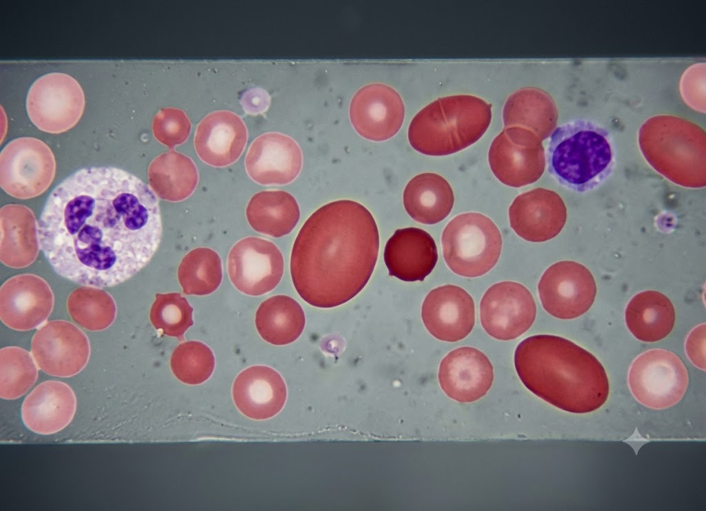
Anisocytosis is a blood test finding that shows your red blood cells aren’t all the same size. Some may be smaller than normal, some larger, and some a mix of both. Doctors usually spot it on a complete blood count (CBC), particularly when the red cell distribution width (RDW) comes back higher than expected. On its own, anisocytosis isn’t a disease it’s a clue pointing toward something else going on in the body.
This size variation can help explain symptoms like fatigue, weakness, shortness of breath, dizziness, or unusual paleness. It’s commonly tied to anemia, nutrient shortages, blood loss, long-term illness, bone marrow issues, or recovery from certain treatments. What it actually means depends on the rest of your bloodwork hemoglobin, MCV, iron levels, B12, folate, and what the cells look like under a microscope. Below are seven common causes and what each one might indicate.
What is Anisocytosis and Why is Red Blood Cell Size Important?
Anisocytosis simply means your red blood cells (erythrocytes) aren’t uniform in size. This matters because consistent cell size is key to how efficiently your blood carries oxygen. When that consistency breaks down, it’s a signal that something is interfering with how red blood cells are being made or matured.
To understand what’s going wrong, it helps to first know what a normal, healthy red blood cell looks like and how its size gets measured.
1. Erythrocyte Structural Dynamics and Functional Requirements
At a basic level, anisocytosis describes unusual variation in the volume of circulating red blood cells. To see why this matters clinically, it helps to look at how a healthy red blood cell is designed. A normal mature erythrocyte measures about 7–8 micrometers across and roughly 2 micrometers thick.
                       [Healthy Red Blood Cell Design]
                                     │
      ┌──────────────────────────────┴──────────────────────────────┐
      ▼                                                             ▼
[Biconcave Disc Shape]                                  [Flexible Outer Membrane]
- Flat center boosts surface area for gas exchange     - Bends without breaking
- Helps oxygen move quickly in and out                 - Fits through 4-micron capillaries
- Holds around 270 million hemoglobin molecules         - Reduces clogging and friction
Rather than being a solid sphere, a healthy red blood cell is shaped like a flattened, dimpled disc closer to a doughnut than a ball. This shape gives it more surface area relative to its size, which speeds up gas exchange, letting the cell pick up oxygen quickly in the lungs and release it efficiently in the body’s tissues.
Each cell carries about 270 million hemoglobin molecules the iron-containing protein responsible for binding oxygen. Because cells are normally uniform in size, each one carries a predictable hemoglobin load, which keeps the blood’s overall oxygen capacity steady.
This same disc shape, paired with a flexible membrane, also makes red blood cells highly bendable. They can twist and squeeze through capillaries as narrow as 4 micrometers without rupturing. When anisocytosis disrupts this uniformity, both blood flow and oxygen delivery can suffer.
2. Laboratory Quantification and Automated Diagnostics
A standard CBC is the main tool used to detect anisocytosis. While measures like hemoglobin tell you the overall red blood cell volume, they don’t reveal whether individual cells vary in size.
[Automated CBC] ──► Measures Size Variation ──► Raises RDW % ──► Signals Anisocytosis
To catch this, lab analyzers calculate the Red Cell Distribution Width (RDW) a statistical measure of how much cell volume varies within a sample.
A healthy RDW typically falls between 11.5% and 15.0%. Anything higher is generally considered the clearest lab sign of anisocytosis, meaning the bone marrow is releasing red blood cells of inconsistent sizes.
Reading RDW Alongside MCV
To pin down the likely cause, doctors compare an elevated RDW against the Mean Corpuscular Volume (MCV), which reflects the average cell size.
| Pattern | RDW | MCV | Likely Cause |
|---|---|---|---|
| Microcytic Anisocytosis | High (>15.0%) | Low (<80 fL) | Early iron deficiency anemia |
| Macrocytic Anisocytosis | High (>15.0%) | High (>100 fL) | B12 or folate deficiency |
| Uniform Microcytosis | Normal (11.5–15.0%) | Low (<80 fL) | Thalassemia trait |
7 Primary Causes of Anisocytosis
The main causes behind anisocytosis are: iron deficiency anemia, vitamin B12 or folate deficiency, thalassemia, myelodysplastic syndromes, chronic liver disease or heavy alcohol use, recent blood transfusions, and chemotherapy. Each one interferes with the bone marrow’s normal red blood cell production in a different way, leading to size inconsistency. Here’s how each plays out.
1. Microcytic Etiologies: Iron Deficiency and Thalassemia
When a CBC shows anisocytosis together with a low MCV, that points to microcytic anisocytosis the bone marrow is putting out cells that are both undersized and inconsistent in size. The two main drivers here are iron deficiency and thalassemia.
                      [Microcytic Anisocytosis Causes]
                                       │
       ┌───────────────────────────────┴───────────────────────────────┐
       ▼                                                               ▼
[Low Iron Levels]                                            [Thalassemia (Genetic)]
- Precursor cells divide extra times                        - Globin chains don't form correctly
- New cells get progressively smaller                       - Extra chains build up in marrow
- Mix of older, normal cells and new tiny ones               - Damaged, irregular cells released
Iron Deficiency Anemia
Iron is essential for building hemoglobin. When iron runs low, the bone marrow can’t fully stock its developing red blood cells (erythroblasts). To compensate, these cells divide more times than usual before maturing, producing smaller cells known as microcytes.
Because this shortage builds up gradually, the effect on cell size isn’t immediate. Early on, there’s still enough iron for near-normal cells; as reserves drop, newer cells get progressively smaller. The result is a bloodstream with both older, normal-sized cells and newer, smaller ones mixed together which is exactly what drives up RDW.
Thalassemia
Thalassemia is an inherited condition where the body under-produces (or fails to produce) one of the globin chains needed to build hemoglobin correctly. This imbalance prevents stable hemoglobin from forming properly.
The leftover, unpaired globin chains build up inside developing red blood cells in the marrow, destroying many before they ever mature. The cells that do survive tend to be small and pale, since they carry less hemoglobin than normal.
This stressed production process leads to a mixed bag of cell types entering circulation. A blood smear from someone with thalassemia often shows a variety of shapes target cells, teardrop-shaped cells, fragmented cells all of which push the RDW higher.
2. Macrocytic Etiologies: Nutritional Deficiencies, Liver Disease, and Chemotherapy
Macrocytic anisocytosis is the flip side a high RDW paired with a high MCV, meaning the circulating cells are both larger than normal and inconsistently sized. This pattern usually points to problems with DNA synthesis or the cell membrane itself.
                      [Macrocytic Anisocytosis Causes]
                                       │
    ┌──────────────────────────────────┼──────────────────────────────────┐
    ▼                                  ▼                                  ▼
[B12 / Folate Shortage]      [Liver Disease / Alcohol Use]      [Chemotherapy]
- DNA replication stalls     - Lipid balance is disrupted        - Stem cell division is disrupted
- Cell body outgrows nucleus - Cholesterol reshapes membranes    - Marrow output becomes erratic
- Oversized cells form       - Cells flatten into larger shapes  - Irregular, large cells released
Vitamin B12 and Folate Deficiencies
B12 and folate are both required for DNA synthesis and cell division. Without enough of either, fast-dividing bone marrow stem cells can’t produce the DNA building blocks they need.
This creates an imbalance called megaloblastic maturation, where the cell’s nucleus lags behind while its cytoplasm keeps growing. The cell keeps expanding but can’t divide the way it normally would.
The bone marrow ends up releasing these oversized, fragile, immature cells (macrocytes) into circulation. Because some cells manage to divide while others don’t, the result is a wide spread of cell sizes and a higher RDW.
Chronic Liver Disease and Alcoholism
The liver helps regulate the cholesterol and fats that make up the red blood cell membrane. When liver function declines as with cirrhosis this regulation breaks down, and excess fat starts depositing into the membranes of circulating cells.
This extra lipid stretches the cell’s surface area relative to its internal volume, producing larger, flattened cells. Since the amount of fat buildup varies from cell to cell, the sizes and shapes end up inconsistent sometimes showing up as target cells or spur cells.
On top of that, heavy alcohol use can directly harm the bone marrow and often causes folate deficiency through poor nutrition, compounding the effect.
Chemotherapy Agents
Chemotherapy drugs are designed to target fast-dividing cells which includes cancer cells, but also healthy, high-turnover tissue like bone marrow stem cells.
This suppresses normal red blood cell production. Chemotherapy drugs interfere with DNA synthesis in a way that mimics B12 or folate deficiency, letting cells grow without dividing properly.
As the marrow tries to recover between treatment rounds, it releases cells at different sizes and stages of development, creating a highly uneven population in the bloodstream.
3. Independent Structural Catalyst Groups: Transfusions and MDS
A few causes of anisocytosis don’t fit neatly into the nutrient-deficiency category they’re either a form of bone marrow disease or a temporary mixing of two different blood populations.
[Patient's Small Cells] ──► [Transfusion of Normal Cells] ──► Mixed Population (High RDW)
Myelodysplastic Syndromes (MDS)
MDS refers to a group of bone marrow disorders where blood cell production becomes disorganized and dysfunctional. Mutated stem cells lose their ability to mature properly into healthy blood cells.
This affects red blood cell development at every stage, producing cells that vary widely in size, shape, and hemoglobin content. Unlike the more predictable pattern seen in nutrient deficiencies, MDS can cause an extreme mix of small, normal, and oversized cells sometimes with unusual features like multiple nuclei pushing RDW very high.
Recent Blood Transfusions
A recent transfusion is a common, temporary cause of anisocytosis. It introduces a second, different population of red blood cells into the bloodstream.
For example, someone with iron deficiency anemia (whose native cells are small) who receives donor blood (with normal-sized cells) will temporarily have both cell types circulating at once.
When a lab analyzer measures this mixed sample, it detects a wide range of cell sizes, raising the RDW. This effect fades over time as the donor cells age out and the underlying anemia is treated.
What Does Anisocytosis Mean for Your Red Blood Cells and Overall Health?
Anisocytosis means your red blood cells are likely less efficient at carrying oxygen, which can bring on anemia-type symptoms. But its real importance lies in what it signals an underlying condition that needs to be identified and treated. It’s a sign of a production problem, not a problem in itself.
1. Impairment of Ocular and Microvascular Gas Exchange
Anisocytosis reflects a structural issue that can reduce how well blood carries oxygen. When cells vary a lot in size, they lose the ideal disc shape built for efficient gas exchange.
                        [Effects of Size Variation]
                                       │
       ┌───────────────────────────────┴───────────────────────────────┐
       ▼                                                               ▼
[Smaller Cells (Microcytic)]                          [Larger Cells (Macrocytic)]
- Carry less hemoglobin                                - Less surface area per volume
- Lower oxygen-carrying capacity                        - Struggle through narrow capillaries
- Can lead to tissue hypoxia                            - Break down (hemolyze) more easily
Smaller cells simply don’t hold enough hemoglobin, which limits how much oxygen they can carry contributing to tissue-level oxygen shortage.
Larger cells, meanwhile, are often structurally weaker. Their fragile membranes rupture more easily, shortening their lifespan and reducing the overall number of healthy cells in circulation. Their bulkier, rounder shape also slows gas exchange and makes it harder for them to pass through tiny capillaries, which can restrict blood flow to organs.
2. Clinical Symptom Matrix of Systemic Hypoxia
The symptoms tied to anisocytosis usually come from the oxygen shortage caused by the underlying condition not from the size variation itself.
[Reduced Oxygen Delivery] ──► Tissue Hypoxia ──► Fatigue & Breathlessness
- Fatigue and low energy muscles and the brain don’t get enough oxygen to keep up with daily demands.
- Shortness of breath with activity the heart and lungs work harder to compensate for lower oxygen-carrying capacity.
- Pale skin, eyes, or nail beds a visible sign of reduced oxygenated blood near the surface.
- Cold hands and feet the body redirects blood flow away from the extremities to protect core organs.
- Heart palpitations in more severe cases, the heart beats faster to circulate the reduced oxygen supply.
3. Pathological Classification: Diagnostic Sign vs. Active Disease
It’s important to understand that anisocytosis isn’t a standalone illness it’s a lab marker that flags a disruption in red blood cell production.
                  [Anisocytosis in Context]
                                     │
     ┌───────────────────────────────┴───────────────────────────────┐
     ▼                                                               ▼
[Think of It Like a Fever]                       [It Prompts Further Testing]
- Signals something is off                        - Flags a marrow production issue
- Doesn't identify the cause alone                - Leads to targeted follow-up tests
- Treating it directly won't fix anything          - Resolves once the root cause is treated
Comparing anisocytosis to a fever is a useful way to think about it a fever tells you something’s wrong without telling you exactly what. Similarly, an elevated RDW flags a disruption in red blood cell production and prompts further investigation.
The real value is in what it points to anything from a simple nutrient deficiency to a more serious bone marrow disorder or liver condition. Treatment is never aimed at the anisocytosis itself, but at whatever is causing it. Once that underlying issue is resolved, the bone marrow goes back to producing uniform cells and the anisocytosis clears up on its own.
The Diagnostic Nuances and Related Conditions of Anisocytosis
Understanding anisocytosis fully means looking closely at RDW, classifying the type of size variation, and distinguishing it from related findings like poikilocytosis (irregular cell shape). This matters because treatment always targets the underlying cause, not the anisocytosis itself.
By reviewing the CBC and a blood smear together, doctors can trace the root cause and figure out the right treatment path.
1. Comprehensive Laboratory Assessment: The Interplay of RDW, MCV, and Reticulocytes
To fully interpret anisocytosis, doctors don’t look at RDW alone they combine it with MCV and the reticulocyte count. Together, these three values show both what the cells look like and how active the bone marrow currently is.
                     [Three Key Lab Values]
                                         │
       ┌─────────────────────────────────┼─────────────────────────────────┐
       ▼                                 ▼                                 ▼
[RDW]                              [MCV]                              [Reticulocyte Count]
- Measures size variation          - Measures average cell size       - Measures new cell output
- Flags anisocytosis               - Sorts into micro/macro/normal    - Shows marrow responsiveness
The Reticulocyte Count: Assessing Bone Marrow Activity
Reticulocytes are newly made, immature red blood cells. They spend roughly a day maturing in the marrow, then finish maturing in the bloodstream over the next 24–48 hours. A normal count sits between 0.5% and 2.5%.
- High reticulocyte count with high RDW: suggests the marrow is actively pushing out new cells to make up for something acute like sudden blood loss or active cell destruction. Since these young cells are physically bigger than mature ones, they raise the RDW.
- Low or normal reticulocyte count with high RDW: suggests a production problem the marrow isn’t able to keep up despite a shortage of healthy cells. This is typical of nutrient deficiencies or conditions like MDS or aplastic anemia.
2. Advanced Microscopic Analysis: Sorting Out Size vs. Shape
While automated analyzers give precise numbers for MCV and RDW, confirming a complex anisocytosis case usually requires a manual look at a blood smear.
                           [Blood Smear Review]
                                         │
       ┌─────────────────────────────────┴─────────────────────────────────┐
       ▼                                                                   ▼
[Anisocytosis: Size]                                [Poikilocytosis: Shape]
- Microcytes (under 6 µm)                            - Schistocytes (fragmented, torn cells)
- Macrocytes (over 8.5 µm)                            - Spherocytes (lost membrane)
- Mixed generations of cells                          - Target cells (excess membrane)
Pathological Identification of Cell Sizes
A pathologist typically compares cell size to a normal small lymphocyte’s nucleus, which is about 8 micrometers, as a reference point.
- Microcytes: under 6 micrometers, often with a widened pale center, signaling low hemoglobin.
- Macrocytes: over 8.5 micrometers. Oval-shaped macrocytes are a classic sign of B12 or folate deficiency.
The Diagnostic Value of Specific Cell Shapes (Poikilocytosis)
When anisocytosis shows up alongside shape changes (poikilocytosis), the specific shapes seen can point to a cause:
- Schistocytes (fragmented, jagged cells): suggest mechanical destruction, often from cells squeezing through damaged small vessels seen in serious conditions like MAHA or DIC.
- Spherocytes (small, dark, round cells with no pale center): occur when part of the membrane is stripped away, often in autoimmune hemolytic anemia or hereditary spherocytosis.
- Target cells: look like a bullseye due to excess membrane relative to hemoglobin content commonly seen in thalassemia or liver-related lipid imbalances.
3. Pathophysiology of Nutrient Deficiencies: Cellular Disruption Mechanics
Nutrient deficiencies cause anisocytosis by interfering with cell development in the marrow. Iron deficiency and B12/folate deficiency do this through very different mechanisms.
                          [Nutrient Deficiency Pathways]
                                         │
       ┌─────────────────────────────────┴─────────────────────────────────┐
       ▼                                                                   ▼
[Iron Deficiency]                                   [B12 / Folate Deficiency]
- Hemoglobin assembly slows                          - DNA replication stalls
- Cells divide an extra time                         - Nucleus lags behind cytoplasm growth
- Results in small, uneven cells                      - Results in large, fragile cells
The Biology of Iron Deficiency Anemia
Normally, developing red blood cells build hemoglobin continuously. When iron is low, hemoglobin assembly slows down. To adjust, the cell divides an extra time, producing smaller cells (microcytes).
Because this happens gradually over weeks or months, the marrow ends up releasing a mix of older, normal-sized cells and newer, smaller ones which drives up RDW.
The Biology of Vitamin B12 and Folate Deficiencies
B12 and folate are both needed to make the building blocks for DNA replication. Without them, DNA synthesis slows, but RNA and protein production in the cytoplasm continue normally.
This mismatch means the nucleus can’t divide on schedule, while the cytoplasm keeps growing producing an oversized cell called a megaloblast. Once these cells enter circulation as macrocytes, they’re larger, more fragile, and shorter-lived, often breaking down prematurely in the spleen.
Conclusion
Anisocytosis is a useful warning sign it shows your red blood cells vary more in size than they should. Common causes include iron deficiency, B12 or folate deficiency, blood loss, hemolytic anemia, chronic illness, and bone marrow disorders. It shouldn’t be interpreted on its own, though it needs to be read alongside your other bloodwork and symptoms. If your test shows anisocytosis, a healthcare provider can help identify the cause and the right next steps.
Frequently Asked Questions
1. What is anisocytosis?
Anisocytosis means your red blood cells show more size variation than normal some smaller, some larger than they should be. It’s usually picked up on a CBC or blood smear. It’s not a diagnosis on its own, but it can point toward anemia or another blood condition.
2. What causes anisocytosis?
It can stem from several conditions affecting red blood cell production or survival, including iron deficiency, B12 or folate deficiency, recent blood loss, hemolytic anemia, and chronic inflammation. It can also show up with certain bone marrow disorders or during anemia treatment. The exact cause depends on other lab results and your medical history.
3. How is anisocytosis detected on a blood test?
It’s usually flagged by an elevated RDW, which measures how much red blood cells vary in size. A blood smear can also show whether cells look uneven, small, large, or mixed. Doctors typically compare RDW with hemoglobin, MCV, iron, B12, and folate levels to understand the full picture.
4. Does anisocytosis always mean anemia?
Not necessarily. It’s common in different types of anemia, since cell size can shift when the body lacks iron, B12, or folate but it can appear before anemia becomes obvious. Your provider will look at the full blood count to determine whether anemia is present.
5. How is anisocytosis treated?
Treatment depends on the underlying cause. Iron deficiency is treated with iron replacement (and identifying why iron is low). B12 or folate deficiency may call for supplements or dietary changes. If it’s linked to chronic disease or bone marrow issues, treatment focuses on managing that underlying condition.