Bronchoscopy Explained: Why It’s Done and What It Can Diagnose
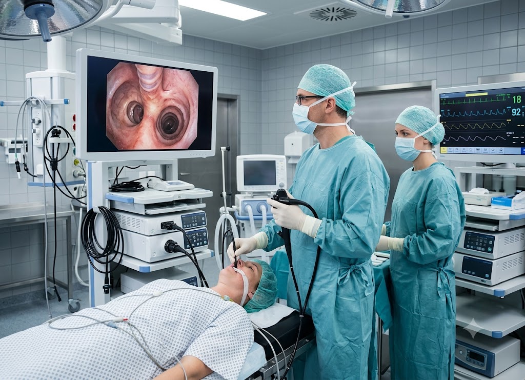
A bronchoscopy is a procedure that lets doctors examine the inside of your airways and lungs using a thin, flexible tube fitted with a light and a tiny camera, called a bronchoscope. The scope is guided through the nose or mouth, down into the windpipe, and into the bronchi, giving your medical team a close-up view of your respiratory tract. Beyond simply looking around, the procedure can also be used to collect tissue samples, clear out mucus or foreign material, and carry out certain treatments. It’s one of the most useful tools available for diagnosing and managing lung conditions.
Your doctor might suggest a bronchoscopy if you’re dealing with a cough that won’t go away, coughing up blood, an unexplained spot on a chest X-ray or CT scan, lung infections that keep coming back, or a suspected lung disease. The procedure can uncover infections, inflammation, tumors, narrowed airways, and other problems that imaging alone often can’t fully explain.
Depending on why the test is being done, your doctor may also take biopsies or collect fluid for lab testing. Below, we’ll walk through why bronchoscopies are performed, what they can diagnose, how to get ready, and what the experience looks like from start to finish.
Why is a Bronchoscopy Performed on the Lungs?
Doctors turn to bronchoscopy for several important reasons: to investigate something unusual on an imaging scan, to work out what’s behind ongoing breathing symptoms, to gather tissue for testing, and in some cases, to physically treat a blockage in the airway.
Think of it as a direct line of sight into the airway system offering real answers where X-rays and CT scans can only raise questions. To fully appreciate its value, it helps to understand what the procedure actually involves and which symptoms typically lead a doctor to order one.
Clinical Indication and Airway Access
A bronchoscopy gives pulmonologists and thoracic surgeons a firsthand look at the trachea and bronchial tubes, helping them evaluate issues that scans alone can’t fully clarify.
There are two main types of bronchoscopes used, depending on the situation:
Flexible Bronchoscopy the more commonly used option. Its thin, bendable design lets the doctor navigate deep into the smaller airway branches. It’s typically done with light sedation and a local numbing agent, which calms the gag reflex while keeping the patient comfortable and relaxed throughout.
Rigid Bronchoscopy this version uses a straight metal tube and requires general anesthesia in an operating room, since it needs a wider path to work through. Its bigger channel makes it the better choice for handling serious bleeding, inserting airway stents, or clearing large blockages.
What is a Bronchoscopy Used to Diagnose?
One of the main purposes of a bronchoscopy is to get to the bottom of unexplained breathing problems or lung changes. Here’s a look at the most common reasons doctors order this test:
Unusual Findings on Chest Imaging If a CT scan or X-ray turns up a nodule, mass, spot, or swollen lymph nodes that can’t be explained, a bronchoscopy is often the next step to inspect the area directly.
Coughing Up Blood (Hemoptysis) This symptom needs prompt attention. A bronchoscopy helps doctors pinpoint exactly where the bleeding is coming from, whether it’s an infection, tumor, or localized inflammation.
A Cough That Won’t Quit When a cough sticks around for more than eight weeks with no obvious explanation like asthma or reflux, doctors may look inside to rule out things like an early tumor, ongoing inflammation, or something inhaled that got stuck.
Wheezing or Stridor in One Spot General wheezing usually points to asthma, but wheezing isolated to one area of the chest can signal a partial blockage. Stridor a high-pitched breathing sound often means the upper airway has narrowed. The scope lets doctors confirm whether it’s a tumor, scar tissue, or mucus causing the issue.
Pneumonia That Keeps Returning to the Same Spot If pneumonia keeps showing up in the exact same area of the lung, there’s usually a structural reason, like something blocking normal drainage. The scope helps doctors check for things like an enlarged lymph node pressing on the airway from outside.
Tissue Collection: The Bronchoscopy Biopsy
If the specialist spots a mass or abnormal-looking area during the exam, they can thread small instruments through the scope to collect samples for closer analysis this is what’s known as a bronchoscopy biopsy.
Forceps Biopsy Tiny forceps pinch off small tissue samples from a visible abnormality along the airway.
Bronchial Brushing A small brush gently gathers cells from the surface lining of the airway.
Bronchoalveolar Lavage (BAL) Sterile saline is flushed into a specific part of the lung, then suctioned back out. The fluid carries cells and microorganisms from deep in the lungs, which is especially helpful for catching hard-to-find infections or inflammation.
These targeted samples give pathologists what they need to determine whether an abnormality is cancerous, infectious, or tied to an autoimmune condition critical information for building your treatment plan.
Navigating Procedure Safety
Is a bronchoscopy dangerous? For most patients, it’s a routine, low-risk outpatient procedure. Some people experience a sore throat, mild coughing, or slight hoarseness afterward as the scope passes near the vocal cords all typically temporary.
More serious risks like a small tear in the lung (pneumothorax) or notable bleeding are uncommon but can happen, especially when a deeper biopsy is taken. Throughout the procedure, your care team keeps a close eye on your oxygen levels, heart rate, and breathing to keep things safe and well-controlled.
What Conditions Can a Bronchoscopy Reveal?
Bronchoscopy can uncover a wide range of lung issues from cancerous tumors and infections like pneumonia or tuberculosis, to inflammatory conditions such as sarcoidosis, airway blockages from mucus or foreign objects, active bleeding, and structural narrowing (stenosis).
Because it allows both direct visualization and tissue sampling, it’s one of the most reliable tools doctors have for confirming a diagnosis with actual cellular evidence not just an educated guess from imaging.
Defining the Scope of Internal Airway Visualization
At its core, a bronchoscopy is an advanced endoscopic procedure that gives a pulmonologist a direct look inside the respiratory system. When a chest scan shows something unexplained, this internal exam is often what’s needed to confirm exactly what’s going on.
Rather than relying on outside guesswork, this real-time camera view gives your care team a precise, physical picture of the trachea and bronchial tubes moving from speculation to solid evidence.
Diagnosing Complex Lung Diseases via Cellular Sampling
A standard bronchoscopy can help identify many different lung diseases by collecting cells and fluid straight from the source:
Infections For patients with pneumonia that isn’t responding to antibiotics, or those with weakened immune systems, a fluid wash (BAL) can isolate the specific pathogen. This is particularly useful for spotting infections like Pneumocystis jirovecii pneumonia (PJP) or confirming tuberculosis when a standard cough sample comes back negative.
Inflammatory and Autoimmune Conditions Diseases like sarcoidosis create small clusters of inflammatory cells called granulomas. A targeted biopsy can collect these clusters and rule out other conditions that might look similar on a CT scan.
Interstitial Lung Diseases When scar tissue builds up progressively in the lungs, a transbronchial biopsy allows doctors to take small samples directly through the airway wall, helping distinguish between conditions like idiopathic pulmonary fibrosis and other forms of pneumonitis.
Detecting and Staging Lung Cancer
Bronchoscopy is one of the best tools for finding, mapping, and evaluating tumors in the central airways.
Direct Visualization and Sampling If a tumor is growing inside the main bronchial walls, the doctor can see it clearly on screen and use forceps or a brush to collect cells for a pathologist to review.
Endobronchial Ultrasound (EBUS) For tumors just outside the airway wall, or to check nearby lymph nodes, doctors use a scope with a built-in ultrasound probe. This lets them see through the airway wall and guide a fine needle precisely into a lymph node to collect cells a process called transbronchial needle aspiration (TBNA). It’s essential for determining whether lung cancer has spread.
Electromagnetic Navigational Bronchoscopy (ENB) For small nodules tucked away in the outer edges of the lungs, this technology builds a 3D map from a prior CT scan, essentially acting as a GPS to guide the biopsy right to the nodule.
Identifying Structural and Mechanical Abnormalities
Bronchoscopy isn’t just for microscopic disease it also helps identify physical problems with the airway itself:
Airway Obstructions The scope reveals exactly what’s causing a blockage, whether it’s a tumor, hardened mucus, or an enlarged lymph node pressing from outside.
Airway Stenosis This is an abnormal narrowing, often from scar tissue caused by long-term breathing tubes or chronic infections. The scope helps doctors gauge how severe the narrowing is and plan treatment, such as a balloon dilation or stent placement.
Tracheobronchomalacia This happens when the cartilage supporting the windpipe becomes weak or floppy. Because the scope shows the airway in real time, doctors can watch it collapse as the patient exhales, confirming the diagnosis directly.
Locating Bleeding Sources When someone is coughing up blood, the scope lets doctors check each airway branch methodically to find the exact source, whether it’s a damaged blood vessel or inflamed tissue.
How is a Bronchoscopy Procedure Conducted?
The process starts with preparation fasting and medication adjustments followed by sedation and numbing medication for comfort, and finally, insertion of the flexible scope to examine the airways and collect samples if needed.
Pre-Procedure Preparation Framework
Good preparation is key to a smooth, low-risk bronchoscopy. Your care team will walk you through several important steps beforehand:
Fasting Beforehand You’ll need to avoid eating or drinking anything, including water, for six to eight hours before the test. Since sedation and numbing sprays suppress your gag reflex, an empty stomach helps prevent aspiration a serious complication where stomach contents enter the lungs.
Reviewing Your Medications Your doctor will go over everything you’re taking. Blood thinners like warfarin, apixaban, clopidogrel, or aspirin are usually paused a few days beforehand to lower bleeding risk, especially if a biopsy is planned. If you have diabetes, you’ll get specific guidance on adjusting insulin or oral medications the morning of the test.
Arranging a Ride Home Sedation will leave you groggy and impair your judgment for hours afterward, so you’ll need someone to drive you home. Avoid driving, operating machinery, or signing anything important for at least 24 hours.
Pre-Test Health Check Before your appointment, you’ll typically have a physical exam, an allergy review, and blood work to check your clotting ability and blood counts, so your care team can tailor the procedure to your needs.
Real-Time Procedure Room Process
The procedure itself happens in an endoscopy suite or specialized treatment room, closely supervised by a pulmonologist, nurses, and sometimes an anesthesiologist.
Getting Set Up You’ll change into a gown, lie down on the procedure bed, and have an IV placed in your arm. Monitors will track your blood pressure, heart rhythm (ECG), and oxygen levels throughout, while a nasal cannula supplies extra oxygen.
Sedation and Numbing A sedative delivered through the IV helps you relax deeply this is called conscious sedation, meaning you’re deeply relaxed but can still breathe independently and respond if needed. A numbing spray, like lidocaine, is then applied to your throat and nose to suppress the gag reflex and reduce coughing.
Inserting the Scope Once you’re comfortable, the doctor guides the thin scope through your nostril or mouth. You may be asked to swallow to help it pass through your throat and vocal cords into the trachea, where the physician examines the airway lining on a monitor in real time.
Targeted Internal Sampling Protocols
If something abnormal shows up during the visual exam, the doctor may collect samples using specialized tools passed through the scope:
Lung Biopsy Tiny forceps snip away small pieces of suspicious tissue, giving a definitive answer on whether it’s cancer or scarring disease.
Bronchoalveolar Lavage (BAL) A saline flush collects fluid, cells, and microbes for lab analysis when infection or inflammation is suspected.
EBUS-Guided Sampling For masses or lymph nodes just outside the airway wall, ultrasound guidance helps direct a fine needle to collect cells critical for cancer staging.
Once everything needed has been gathered, the scope is gently withdrawn. The whole process usually takes 30 to 60 minutes, depending on complexity.
Addressing Patient Safety and Risks
How dangerous is a bronchoscopy, really? It’s considered a safe, standard outpatient test. Serious complications are rare, thanks to continuous monitoring of your vitals and oxygen levels throughout.
Minor effects a sore throat, light cough, or blood-tinged spit are common and usually clear up within a few days. Rarer risks, such as a small lung tear or significant bleeding, are more closely tied to deeper biopsies than to the scope itself, making bronchoscopy a precise and generally low-risk way to evaluate lung health.
Overview of the Bronchoscopy Journey
What is a Bronchoscopy A safe procedure using a thin, camera-equipped tube to look directly inside the lungs’ airways.
Bronchoscopy Definition An endoscopic method for examining the respiratory tract, clearing blockages, and collecting cell samples.
What is Bronchoscopy Used for Investigating abnormal scans, tracking down the source of coughing or bleeding, and performing targeted biopsies.
The Risks and Recovery After a Bronchoscopy
While generally safe, bronchoscopy does carry some risk mainly a sore throat and mild cough, with rare complications like bleeding or a collapsed lung. Recovery typically involves a few hours of monitoring at the facility while sedation wears off, followed by a restful day at home.
Post-Procedure Recovery Timeline
Immediate Monitoring After the scope is removed, you’ll be moved to a recovery area where nurses track your heart rate, blood pressure, breathing, and oxygen levels for one to two hours as sedation fades. If a deeper biopsy was performed, a chest X-ray may be taken to confirm your lungs are fully inflated.
Waiting for Your Gag Reflex to Return Since your throat was numbed, you can’t eat or drink right away. A nurse will check that your gag reflex has returned before you’re offered ice chips or sips of water, followed by a light meal.
Going Home Once you’re alert, stable, and able to swallow safely, you’ll be discharged to your pre-arranged ride.
At-Home Recovery Care Guidelines
Take It Easy Residual sedation can affect your reaction time and judgment, so avoid driving, operating machinery, or signing important documents for at least 24 hours. Plan to rest for the remainder of the day.
Managing Mild Discomfort A scratchy throat, light cough, or low-grade fever in the first 24 hours is normal and simply your body’s response to the procedure. Lozenges and over-the-counter pain relief can help.
Getting Back to Normal Most people resume their usual diet, medications, and routine the next day. Your doctor’s office will follow up on results while some initial observations may be shared right after the test, full biopsy or culture results usually take three to seven days.
Addressing Risks: How Dangerous is a Bronchoscopy?
The short answer: it’s a safe, routine outpatient test, though like any procedure, it carries some risk depending on your health and whether a biopsy was taken.
Common, Mild Side Effects These typically resolve within 48 hours on their own:
- A sore or scratchy throat, or mild hoarseness from the scope passing the vocal cords
- A mild cough, sometimes with small streaks of blood, especially after a biopsy
- A brief low-grade fever in the first 24 hours
Rare but Serious Complications These need prompt medical attention:
- Significant Bleeding Minor oozing after a tissue sample is expected, but heavier bleeding is rare and can be managed during the procedure with cold saline or medication.
- Pneumothorax (Collapsed Lung) Mostly linked to transbronchial biopsies, where a puncture in the thin lung lining lets air escape into the chest cavity. A small chest tube may be needed to fix this.
- Airway Spasms or Breathing Trouble Patients with severe COPD or asthma may experience temporary airway tightening or dips in oxygen. Rescue medications are kept on hand just in case.
- Infection There’s a small risk of introducing bacteria into the lungs, though strict sterile technique keeps this risk low.
When to Seek Immediate Help Contact your doctor or go to the ER right away if you experience:
- Sudden, severe chest pain or worsening shortness of breath
- Coughing up more than a few tablespoons of bright red blood
- A persistent fever above 101°F (38.3°C)
Overview of the Bronchoscopy Experience
What is a Bronchoscopy A safe test using a thin, flexible, camera-equipped tube to view the airways directly.
Bronchoscopy Definition An endoscopic exam of the respiratory tract that also allows blockage removal and cell sample collection.
What is Bronchoscopy Used to Diagnose Investigating suspicious scan findings, tracking bleeding sources, and collecting tissue to check for cancer or infection.
The Different Types and Applications of Bronchoscopy
Bronchoscopy isn’t one-size-fits-all it spans everything from routine flexible exams to advanced ultrasound-guided and therapeutic techniques. This range lets pulmonologists match the approach to the clinical need, whether that’s a simple look-around, detailed sampling, or direct treatment of a blockage.
What was once purely a diagnostic tool has grown into a cornerstone of interventional pulmonology, offering less invasive treatment options across many lung conditions.
Advanced Modalities: Flexible vs. Rigid Interventions
Specialists choose between two main instrument types depending on whether the goal is diagnosis or urgent intervention:
Flexible Bronchoscopy The standard choice for most evaluations, using a bendable tube that reaches deep into smaller airways. Performed with local anesthesia and light sedation, it’s typically used for sample collection and routine biopsies.
Rigid Bronchoscopy A straight, unbending steel tube used under general anesthesia in an operating room. While it can’t reach deep into the lungs, its wide channel supports larger tools making it the go-to for serious bleeding, major blockages, or stent placement.
Precision Diagnostics: Endobronchial Ultrasound (EBUS)
For central lung diseases or cancer staging, standard scopes have limits they can only see the inside surface of the airway. EBUS solves this by combining a flexible camera with a miniature ultrasound probe.
This lets doctors see through the airway wall to evaluate nearby structures, like the lymph nodes in the chest’s center. Using real-time ultrasound, a fine needle is guided directly into a target mass a process called TBNA. Key benefits include:
- Reliable Cancer Staging EBUS is considered the gold standard for determining whether lung cancer has spread, guiding decisions between surgery, chemo, or radiation.
- A Gentler Alternative Before EBUS, reaching deep lymph nodes required an invasive surgery called mediastinoscopy. EBUS achieves the same goal as an outpatient procedure, with a quicker recovery and fewer risks.
- Greater Precision Real-time imaging helps doctors place the needle accurately, improving sample quality while protecting nearby blood vessels. It’s also effective for diagnosing non-cancerous conditions like sarcoidosis.
Therapeutic Interventions in Pulmonology
Bronchoscopy isn’t limited to diagnosis it’s also a treatment tool, letting interventional pulmonologists address serious airway issues without open surgery:
- Clearing the Airway Removing foreign objects (common in kids), suctioning mucus plugs causing lung collapse, or clearing blood clots.
- Shrinking Tumors For tumors blocking an airway, doctors can use lasers, electrocautery, or cryotherapy to destroy the tissue.
- Placing Stents A flexible mesh tube can be inserted to hold a narrowed airway open, similar to a stent used in blood vessels.
- Stopping Bleeding Doctors can apply iced saline, epinephrine, or heat therapy directly at the bleeding source.
Comparing Internal Bronchoscopy vs. External Needle Biopsy
When a scan reveals a suspicious nodule, doctors choose between two sampling methods based on its location.
The Internal Route: Bronchoscopy This approach travels through the body’s natural airways, making it ideal for central lesions near the major bronchial tubes and it’s the only non-surgical way to sample chest lymph nodes via EBUS. Because it stays inside the airway, it generally uses light sedation and carries a lower risk of a collapsed lung.
The External Route: CT-Guided Needle Biopsy Also called a transthoracic needle aspiration (TTNA), this method inserts a needle through the chest wall, guided by real-time CT imaging. It’s the standard for peripheral lesions nodules in the outer lung that a bronchoscope can’t reach. Because the needle punctures the lung lining from outside, it carries a higher risk of a temporary collapsed lung and is usually done with local numbing at the skin.
Summary of Diagnostic and Structural Care
What is Bronchoscopy A safe procedure using a thin, camera-equipped tube to view the respiratory tract directly.
Bronchoscopy Biopsy Passing tools through the scope to collect fluid or tissue, essential for diagnosing infections, scarring, or cancer.
How Dangerous is a Bronchoscopy A safe, routine outpatient test with a low risk of serious complications, making it a dependable way to assess lung health.
Conclusion
A bronchoscopy plays an important role in examining the airways, diagnosing lung conditions, and delivering certain treatments without the need for major surgery. It often provides answers when imaging tests alone can’t explain a patient’s symptoms.
While generally safe, it’s worth discussing the benefits, risks, and recovery details with your healthcare provider ahead of time. Understanding why the procedure is recommended can help you feel more prepared and better appreciate its role in diagnosing and treating lung and airway diseases.
Frequently Asked Questions
1. What is a bronchoscopy?
A bronchoscopy is a medical procedure that uses a thin, flexible, camera-equipped tube to examine the airways and lungs. It’s inserted through the nose or mouth and guided into the windpipe and bronchi. During the test, doctors can inspect the airways, collect samples, and sometimes perform treatment. It’s widely used to diagnose and evaluate lung conditions.
2. Why would someone need a bronchoscopy?
Doctors often order this test to investigate a persistent cough, coughing up blood, unexplained shortness of breath, or abnormal imaging results. It also helps diagnose infections, lung cancer, inflammatory disease, or airway blockages, and can be used to monitor known conditions or check how treatment is progressing.
3. Is a bronchoscopy painful?
Not typically the throat is numbed and most patients receive sedation to stay relaxed. You might feel pressure or an urge to cough as the scope moves through, but significant pain is rare. A mild sore throat, hoarseness, or cough may linger for a day or two afterward, and your care team monitors you until sedation wears off.
4. What can a bronchoscopy diagnose?
It can help identify lung cancer, infections, airway inflammation, sarcoidosis, tuberculosis, and other lung diseases, along with airway narrowing, bleeding, inhaled objects, or abnormalities seen on scans. Biopsies, washings, or fluid samples collected during the test help confirm a diagnosis and guide treatment.
5. How should I prepare for a bronchoscopy?
Your provider will give you guidance on eating, drinking, and medications beforehand most people are asked to fast for several hours to lower aspiration risk during sedation. Be sure to mention any blood thinners, allergies, or existing conditions, and arrange for someone to drive you home afterward.
6. What are the risks of a bronchoscopy?
It’s generally a safe procedure with uncommon serious complications. Possible risks include bleeding, infection, sedation reactions, low oxygen, or a collapsed lung particularly with a biopsy. Most side effects, like a sore throat or mild cough, clear up within a few days. Your doctor will talk through the specific risks and benefits based on your health and the reason for the test.