7 Causes of Hyperinflated Lungs and What They Mean
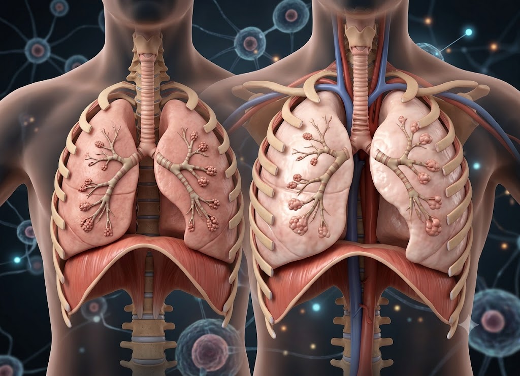
Hyperinflated lungs occur when air becomes trapped inside the lungs, causing them to expand more than they normally should. Because the lungs cannot empty completely, there may be less space available for fresh air during the next breath.
Some people discover they have hyperinflated lungs after having a chest X-ray or CT scan, while others may experience symptoms such as shortness of breath, chest tightness, wheezing, or decreased exercise tolerance. Although the term can sound concerning, it generally indicates that doctors should investigate whether an underlying breathing condition is present.
Knowing the possible causes can make this finding easier to understand. COPD, emphysema, asthma, cystic fibrosis, chronic bronchitis, small airway disorders, and long-term air trapping can all contribute to lung hyperinflation. In some cases, imaging may show hyperinflation before noticeable symptoms develop, while in others it occurs alongside an already diagnosed lung condition. This article explores seven causes of hyperinflated lungs and what they can indicate about airflow and lung function.
Defining Hyperinflation: The Structural Impact of Trapped Air
To understand what is hyperinflation of the lungs, it helps to first understand how air normally moves through the respiratory system. Hyperinflation of the lungs is a condition in which an unusually large amount of used air remains trapped in the respiratory system after normal exhalation. Because that stale air remains inside, there is less room for fresh, oxygen-rich air to enter.
Hyperinflation of Lungs Meaning: The Mechanical Breakdown
To better understand the hyperinflation of lungs meaning, consider the balance between two natural forces. The lungs naturally tend to recoil inward, while the chest wall tends to move outward. Under normal circumstances, these forces remain balanced at the end of a relaxed breath.
When the lungs are hyperinflated, this balance changes. Lung tissue may lose some of its natural elasticity and become less capable of recoiling after inhalation. As a result, the lungs can remain overstretched and hold onto excess air.
Key Pulmonary Volumes
Pulmonary Function Tests (PFTs) can measure important lung volumes and help doctors identify air trapping:
Elevated Functional Residual Capacity (FRC): This refers to the amount of air remaining in the lungs after a normal, relaxed exhalation. When FRC is elevated, more stale air is taking up space that could otherwise be used for fresh air.
Elevated Residual Volume (RV): This is the amount of air left in the lungs after a person exhales as forcefully as possible. A high RV suggests that a significant amount of air remains trapped and cannot be expelled completely.
Altered RV/TLC Ratio: The Residual Volume compared with Total Lung Capacity ($TLC$) helps doctors evaluate the extent of air trapping. A higher ratio means that a greater portion of total lung volume consists of air that remains trapped.
Physiological Signs and Symptoms of Chronic Air Trapping
The mechanical effects of pulmonary hyperinflation can lead to noticeable symptoms and, over time, changes in the shape and function of the chest.
[Loss of Lung Elastic Recoil] ──► [Stale Air Trapped in Alveoli] ──► [Diaphragm Flattens] ──► [Barrel Chest Formed]Chronic Shortness of Breath (Dyspnea): Shortness of breath is one of the most common symptoms associated with hyperinflated lungs. When the lungs already contain trapped air, taking a deep, comfortable breath can become difficult. Breathing may feel shallow, and breathlessness can become more noticeable during even modest physical activity.
Development of a Barrel Chest: Long-term outward pressure from hyper inflated lungs can cause the rib cage to remain expanded and develop a rounded appearance often described as a barrel chest.
Diaphragmatic Flattening: Normally, the diaphragm is a dome-shaped muscle that moves downward and upward to support breathing. Hyperinflation of the lungs can push this muscle downward and flatten it. This reduces its mechanical efficiency and may cause the body to depend more heavily on muscles in the neck and chest, making breathing more tiring.
Persistent Wheezing and Chest Tightness: When air moves through narrowed or obstructed airways, it can create a high-pitched whistling sound known as wheezing. Airway resistance may also cause a continuing sensation of tightness or pressure across the chest.
Reduced Exercise Tolerance: When breathing requires more effort, physical activity can become increasingly difficult. Inefficient airflow and gas exchange may contribute to faster fatigue and reduced physical stamina.
Clinical Identification and Differentiating Types
When someone asks, “should i worry about hyperinflated lungs?” the answer depends on the underlying cause and whether the air trapping is permanent or occurs mainly during activity.
                              [Classification of Lung Hyperinflation]
                                               │
          ┌────────────────────────────────────┴────────────────────────────────────┐
          ▼                                                                         ▼
 [Static Hyperinflation]                                                   [Dynamic Hyperinflation]
  ├── Etiology: Permanent loss of tissue elasticity                             ├── Etiology: Faster breathing during exercise
  ├── Mechanics: Structural damage to alveoli                                   ├── Mechanics: Next breath begins before full exhalation
  └── Diagnostic: Visible on resting chest X-rays                               └── Diagnostic: Assessed during exercise testing
Static Hyperinflation (Structural Damage)
Static hyperinflation results from permanent structural changes within the lung tissue. It remains present even when a person is resting. When the elastic fibers supporting the air sacs are damaged, the lungs may remain stretched and enlarged. These changes can often be seen on a standard chest X-ray.
Dynamic Hyperinflation (Exertional Air Trapping)
Dynamic hyperinflation develops when breathing becomes faster, commonly during exercise or periods of increased respiratory demand. A person with narrowed airways may start the next inhalation before completely finishing the previous exhalation.
As a result, additional air becomes trapped with each breath. This can produce sudden or severe breathlessness during activity, but the hyperinflation may decrease once the breathing rate slows and the person returns to rest.
Radiographic Findings and Terminology
When a radiologist examines a chest X-ray or CT scan, certain structural features may suggest over-inflated lungs. A report may use terms such as pulmonary hyperaeration or pulmonary hyperinflation to describe the appearance.
| Radiographic Sign | Anatomical Appearance on Scan | Clinical Meaning |
|---|---|---|
| Flattened Diaphragmatic Hemidomes | The normally curved diaphragm looks flatter and is positioned lower than expected. | Suggests significant air trapping that is placing pressure on the main breathing muscle. |
| Increased Retrosternal Clear Space | A larger-than-usual dark area is visible behind the sternum and in front of the heart on a side-view X-ray. | Suggests that the front portions of the upper lungs are expanded. |
| Rib Hyper-Extension | More than 9 to 10 posterior ribs may be visible above the diaphragm, with the ribs appearing more horizontal. | Indicates expansion of the chest wall to accommodate increased lung volume. |
| Widened Intercostal Spaces | The spaces between the ribs appear wider than normal. | Reflects ongoing outward pressure from enlarged lungs. |
7 Primary Causes of Air Trapping in the Lungs
The seven main causes of air trapping include common obstructive lung diseases such as Chronic Obstructive Pulmonary Disease (COPD), emphysema, chronic bronchitis, and asthma, as well as conditions such as cystic fibrosis, bronchiectasis, and Alpha-1 Antitrypsin Deficiency.
Although these disorders have different causes, they can all interfere with the normal movement of air out of the lungs. Airflow may be restricted by swollen airway walls, excessive mucus, tightening of airway muscles, or loss of the lungs’ natural elastic recoil.
Identifying the specific reason for air trapping is important because each condition may require a different treatment approach. The resulting hyperinflation may look similar, but the biological process behind it can vary considerably.
Pathophysiology of Air Trapping
Air trapping occurs when damaged or narrowed airways prevent the lungs from emptying normally during exhalation. As air accumulates, the lungs become hyperinflated and the increased volume can place additional stress on the respiratory system.
When considering hyperinflated lungs causes, the underlying problem may involve a structural or mechanical obstruction somewhere within the respiratory tract. During inhalation, the airways naturally become wider, allowing air to move past certain obstructions. During exhalation, however, the airways naturally become narrower.
When disease further narrows these passages, they may close too early and trap stale air inside the alveoli. This process, also called pulmonary hyperinflation, can force the person to breathe at a higher lung volume and make breathing less efficient.
Common Obstructive Lung Diseases (Causes 1–4)
Obstructive lung disorders are among the most common reasons for chronic air trapping. These diseases interfere with the ability to move air out efficiently, meaning the lungs are hyperinflated even at rest in some cases.
Chronic Obstructive Pulmonary Disease (COPD)
COPD is a progressive inflammatory lung disease and a major cause of chronic hyperinflation of the lungs. It can involve narrowing of the small airways along with damage to the supporting lung tissue. During exhalation, weakened airways may collapse too early, leaving air trapped deep within the lungs and contributing to a persistently expanded chest.
Emphysema
Emphysema permanently damages the thin walls separating the tiny air sacs, or alveoli. This produces two major mechanical problems:
- Loss of Surface Area: Small, efficient air sacs may merge into larger, less effective spaces, reducing the surface available for gas exchange.
- Loss of Elastic Recoil: Damage to the elastic fibers reduces the lung’s natural ability to recoil and push air outward. Without this recoil, air becomes more difficult to expel, contributing to severe hyperinflation of lungs.
Chronic Bronchitis
Chronic bronchitis is associated with a persistent productive cough and can contribute to pulmonary hyperaeration by narrowing the airways. Long-term inflammation causes the airway lining to swell, while increased mucus production can create thick mucus plugs. Together, swelling and mucus make it harder for air to leave the lungs during exhalation.
Asthma
Asthma involves ongoing airway inflammation and episodes of tightening of the smooth muscles surrounding the airways, known as bronchoconstriction. During an asthma flare, the airways can become significantly narrower and trap air beyond the obstruction.
Acute hyperinflation of the lungs may improve after effective treatment, including a rescue inhaler. However, severe or poorly controlled asthma can produce longer-term airway changes that may contribute to persistent over-inflation.
Genetic and Structural Conditions (Causes 5–7)
In addition to COPD and asthma, several other disorders can produce significant air trapping by affecting the airways or lung tissue.
                         [Alternative Air Trapping Pathways]
                                      │
          ┌───────────────────────────┼───────────────────────────┐
          ▼                           ▼                           ▼
 [Cystic Fibrosis (CF)]       [Bronchiectasis Damage]     [Alpha-1 Antitrypsin Def.]
 - Inherited chloride problem - Permanently widened airways - Reduced protective protein
 - Thick, sticky mucus        - Mucus collects in pockets  - Elastase damages air sacs
 - Physical airway plugs      - Repeated infection cycle  - Can cause early emphysema
Cystic Fibrosis (CF)
Cystic Fibrosis is an inherited condition that affects how salt and water move through epithelial cells. In the lungs, this can result in unusually thick, sticky mucus that the cilia cannot clear effectively.
The mucus can form plugs inside the airways and contribute to significant air trapping. Because stagnant mucus can also encourage repeated bacterial infections, ongoing inflammation may worsen lung damage over time.
Bronchiectasis
Bronchiectasis is a chronic condition in which the larger airways become permanently widened, scarred, and structurally distorted. It may develop after serious or repeated infections or in association with certain immune disorders.
Because the damaged airways cannot clear mucus normally, secretions can collect in the widened areas. This can contribute to chronic obstruction, repeated infections, and abnormal lung inflation.
Alpha-1 Antitrypsin Deficiency (AATD)
Alpha-1 Antitrypsin Deficiency is an inherited condition in which the body does not produce enough alpha-1 antitrypsin ($AAT$), a protein that helps protect lung tissue.
The Protective Mechanism of AAT:
In healthy lungs, AAT helps protect delicate lung tissue from neutrophil elastase, an enzyme released by white blood cells as part of normal inflammation. When AAT levels are too low, elastase can damage the elastic fibers within the alveoli. This loss of elastic support can result in a form of emphysema and severe hyperinflated lungs, sometimes appearing at a relatively young age.
Summary Table of Air Trapping Mechanisms
| Primary Etiology | Underlying Anatomical Cause | Primary Mechanism of Air Trapping | Typical Clinical Onset |
|---|---|---|---|
| COPD | Combination of airway inflammation and tissue damage. | Small airways can collapse too early during exhalation. | More common in middle-aged and older adults; strongly associated with smoking history. |
| Emphysema | Destruction of alveolar walls and supporting structures. | Loss of elastic recoil, reducing the lungs’ ability to naturally empty. | Usually develops gradually and is a major component of tobacco-related COPD. |
| Chronic Bronchitis | Enlarged mucus glands and swelling of the airway lining. | Airway blockage from swelling and thick mucus plugs. | Associated with a chronic, recurrent productive cough and irritant exposure. |
| Asthma | Reversible bronchospasm and airway inflammation. | Variable airway narrowing caused by inflammation and muscle tightening. | Often starts in childhood but can occur at any age; may be triggered by allergens or exercise. |
| Cystic Fibrosis | Genetic abnormality affecting epithelial chloride channels. | Thick secretions become dehydrated and block the airways. | Often begins early in life and can affect several organ systems. |
| Bronchiectasis | Permanent widening and damage to bronchial walls. | Impaired mucus clearance allows secretions to collect in damaged airways. | Can occur at any age and may follow serious or repeated infections. |
| AATD Deficiency | Genetic shortage of protective alpha-1 antitrypsin. | Uncontrolled enzyme activity damages elastic fibers in the alveoli. | May appear earlier in adulthood and can affect non-smokers. |
Pharmacological Management: Deflating the Airways
Because hyperinflated lungs are generally the result of restricted airflow during exhalation rather than a disease by themselves, medication is often aimed at opening narrowed airways and controlling inflammation. Increasing airway diameter can improve airflow out of the lungs, helping reduce trapped air and excessive lung expansion.
                           [Pharmacological Interventions]
                                      │
          ┌───────────────────────────┴───────────────────────────┐
          ▼                                                       ▼
 [Bronchodilator Therapy]                                [Anti-Inflammatory Therapy]
  ├── Beta-2 Agonists (SABA/LABA): Relax airway muscles    ├── Inhaled Corticosteroids (ICS): Reduce inflammation
  └── Anticholinergics (SAMA/LAMA): Reduce constriction   └── Impact: Helps control swelling and flare-ups
Bronchodilators
Bronchodilators are an important part of treatment for hyperinflated lungs causes such as COPD and asthma. They work by relaxing the smooth muscles surrounding the airways, allowing the breathing passages to remain more open.
Beta-2 Agonists: These medicines stimulate beta receptors and relax airway muscles. Short-Acting Beta-Agonists (SABAs), such as albuterol, can provide rapid relief during acute symptoms, while Long-Acting Beta-Agonists (LABAs), such as salmeterol, provide longer-lasting airway relaxation for maintenance treatment.
Anticholinergics / Muscarinic Antagonists: These medications block acetylcholine, a chemical signal involved in airway narrowing. Short-Acting Muscarinic Antagonists (SAMAs), such as ipratropium, and Long-Acting Muscarinic Antagonists (LAMAs), such as tiotropium, are used in different treatment settings. LAMAs are particularly important in COPD management and provide sustained bronchodilation.
Corticosteroids
When persistent inflammation causes airway swelling and increased mucus production, hyperinflation of the lungs may become worse. Inhaled Corticosteroids (ICS), including fluticasone and budesonide, act locally in the airways to reduce inflammation and limit mucus buildup.
In more advanced obstructive lung disease, an ICS may be combined with a LABA or LAMA in a single inhaler. Systemic corticosteroids, including oral or intravenous forms, are generally used for shorter periods during acute respiratory exacerbations.
Advanced Non-Pharmacological and Rehabilitation Strategies
Although medication can help open the airways, non-drug approaches can also help the body cope more efficiently with trapped air and improve breathing mechanics.
Pulmonary Rehabilitation
Pulmonary rehabilitation is a structured program supervised by healthcare professionals. It can combine individualized exercise training, nutritional guidance, and education about managing lung disease.
The exercise component helps improve physical conditioning and strengthen respiratory and supporting muscles. By helping the muscles use oxygen more efficiently, rehabilitation can reduce the breathing demand associated with everyday activities and make breathlessness easier to manage.
Re-Education of Breathing Mechanics
Specific breathing techniques can be useful for managing pulmonary hyperinflation.
Essential Breathing Techniques:
- Pursed-Lip Breathing:
Inhale through nose (2s) ──► Purse lips ──► Exhale slowly & steadily (4s)Slowly breathing out through partially closed lips creates gentle back-pressure in the airways. This can help keep smaller airways open longer during exhalation, allowing more trapped air to leave.- Diaphragmatic (Belly) Breathing: This technique encourages more effective use of the diaphragm during breathing. By focusing on abdominal movement, patients may reduce their reliance on the neck and upper-chest muscles that can become fatigued with chronic breathing difficulty.
Long-Term Oxygen Therapy (LTOT)
In advanced lung disease where oxygen levels in the blood are significantly reduced, physicians may prescribe supplemental oxygen. Oxygen therapy does not directly remove trapped air or reverse structural hyperinflation, but it can help maintain adequate oxygen levels.
In appropriate patients, this may reduce strain on the cardiovascular system, help limit complications related to low oxygen levels, and support physical activity.
Surgical and Bronchoscopic Interventions
When someone asks, “should i worry about hyperinflated lungs?” severe cases require attention to how excessive lung volume affects the rest of the chest. In advanced emphysema, severely damaged parts of the lung may expand into large air-filled spaces known as bullae. These areas can compress healthier tissue and push the diaphragm downward.
For carefully selected patients, procedures may be used to reduce the volume of these severely damaged areas.
                     [Advanced Volume Reduction Techniques]
                                      │
          ┌───────────────────────────┴───────────────────────────┐
          ▼                                                       ▼
 [Lung Volume Reduction Surgery (LVRS)]             [Bronchoscopic Endobronchial Valves (EBV)]
 - Removes severely damaged lung tissue               - Minimally invasive catheter-based approach
 - Often targets the most affected upper areas        - Places one-way valves in selected airways
 - Helps restore diaphragm positioning                 - Prevents new air entering while allowing air out
 - Gives healthier lung regions more room              - Encourages controlled collapse of target tissue
Lung Volume Reduction Surgery (LVRS)
LVRS is a surgical procedure in which a thoracic surgeon removes the most severely damaged and hyperinflated portions of lung tissue, often involving the upper lobes.
Removing these poorly functioning areas can create additional space inside the chest. The remaining healthier lung tissue may then expand more effectively, while the flattened diaphragm can move toward a more useful dome-shaped position.
Bronchoscopic Lung Volume Reduction (BLVR)
For selected patients who are not suitable candidates for open surgery, BLVR can provide a less invasive option. A pulmonologist uses a flexible bronchoscope to place tiny one-way endobronchial valves (EBVs) into airways supplying the most hyperinflated areas.
The valves prevent new air from entering the targeted damaged regions while allowing trapped air and mucus to leave. Over time, the selected non-functioning area may collapse in a controlled manner, reducing pressure on the remaining lung without requiring a surgical incision.
Comprehensive Management Protocol Matrix
| Strategy Class | Specific Intervention | Direct Physiological Impact | Ultimate Clinical Target |
|---|---|---|---|
| Pharmacological | LAMA / LABA Inhalers | Relax airway smooth muscle and help prevent early small-airway closure. | Improves expiratory airflow and may reduce Residual Volume ($RV$). |
| Pharmacological | Inhaled Corticosteroids | Reduces inflammation and limits excessive mucus production. | Helps maintain airway openness and reduce respiratory flare-ups. |
| Rehabilitative | Pursed-Lip Breathing | Creates gentle expiratory back-pressure that supports airway stability. | Encourages more complete exhalation and can reduce dynamic air trapping. |
| Rehabilitative | Pulmonary Conditioning | Improves how effectively skeletal muscles use available oxygen. | Reduces respiratory demand and improves exercise tolerance. |
| Interventional | Endobronchial Valves | Encourages controlled collapse of severely hyperinflated lung regions. | Reduces pressure on the diaphragm and gives functioning lung tissue more room. |
Advanced Diagnostic and Comparative Aspects of Lung Hyperinflation
Understanding lung hyperinflation may require detailed pulmonary function testing and imaging. These tests can help distinguish hyperinflation from opposite conditions such as atelectasis and determine whether air trapping occurs at rest or mainly during activity.
These diagnostic tools and management strategies give healthcare professionals a way to assess the extent of hyperinflation while helping patients learn techniques that may make everyday breathing more manageable.
Advanced Diagnostic Modalities and PFT Metrics
A complete assessment of hyperinflated lungs generally combines Pulmonary Function Tests (PFTs) with chest imaging. Standard spirometry can demonstrate airflow limitation, but it does not directly measure all trapped air volumes. More advanced testing, such as body plethysmography, can provide a more complete measurement of lung volumes.
                         [Advanced Diagnostic Flowchart]
                                      │
          ┌───────────────────────────┴───────────────────────────┐
          ▼                                                       ▼
 [Body Plethysmography]                                  [Expiratory HRCT Scans]
  ├── Measures FRC & RV                                    ├── Shows lung tissue damage
  ├── Assesses airway resistance                           ├── Identifies retained air
  └── Measures thoracic gas volume                         └── Helps detect bullous disease
Body Plethysmography
Whole-body plethysmography is an important clinical test for assessing hyperinflation of the lungs. During the test, the patient sits inside an airtight chamber and breathes through a specialized mouthpiece.
The equipment measures small changes in cabin and mouth pressure to calculate the Absolute Thoracic Gas Volume (VTG). This makes it possible to measure air that cannot be fully exhaled.
Elevated RV and an increased RV/TLC ratio can provide strong evidence of air trapping and help confirm what is hyperinflation of the lungs.
High-Resolution Computed Tomography (HRCT)
A standard chest X-ray can reveal general signs such as a flattened diaphragm or expanded rib cage, but an HRCT scan provides much greater detail of the lung structures.
To investigate hyperinflated lungs causes, doctors may use CT imaging during both maximum inhalation and maximum exhalation.
During expiration, healthy lung tissue becomes denser as air leaves. Areas affected by pulmonary hyperaeration may remain unusually dark because they retain air instead of collapsing normally. This can help identify emphysema, bullous changes, and small-airway obstruction.
Physiological Mechanics: Static vs. Dynamic Hyperinflation
The terms hyper inflated lungs and pulmonary hyperinflation can describe different physiological processes depending on whether excess air remains trapped at rest or accumulates during increased activity.
                    [Mechanical Divergence of Lung Volumes]
                                      │
          ┌───────────────────────────┴───────────────────────────┐
          ▼                                                       ▼
 [Static Hyperinflation (At Rest)]                    [Dynamic Hyperinflation (In Motion)]
 - Caused by reduced elastic recoil                    - Caused by faster breathing
 - Structural damage to alveolar walls                 - Next breath begins too soon
 - Trapped air remains at rest                         - Air progressively "stacks" with each breath
Static Hyperinflation
Static hyperinflation is present even when a person is resting. It is associated with an elevated resting Functional Residual Capacity ($FRC$).
One major cause is significant loss of lung elastic recoil. In conditions such as emphysema, damage to the elastic framework of the alveoli reduces the lungs’ ability to recoil naturally. As a result, more air remains inside after a normal breath, and the chest can remain expanded.
Dynamic Hyperinflation
Dynamic hyperinflation develops when breathing becomes faster, such as during exercise or an acute respiratory illness.
When narrowed airways limit the time available for exhalation, the person may begin another breath before completely emptying the previous one. This expiratory time limitation causes additional air to build up with each breathing cycle.
As the trapped volume increases, the resting lung volume rises, the diaphragm becomes flatter, and severe breathlessness may develop during physical exertion.
Comparative Pathologies: Hyperinflation vs. Atelectasis
Hyperinflation and atelectasis are essentially opposite conditions when considering lung volume, air content, and chest mechanics.
Hyperinflation: Pathological Over-Distension
With hyperinflation, the lungs become excessively expanded because air remains trapped. This results in a pathological increase in Residual Volume (RV) and Total Lung Capacity (TLC).
The underlying problem is usually restricted expiratory airflow. Air may enter the alveoli relatively easily but have difficulty leaving during exhalation.
On a chest X-ray, hyperinflation can appear as enlarged, darker lung fields, wider spaces between the ribs, and a flattened diaphragm.
Atelectasis: Structural Volume Loss
Atelectasis is the opposite process. It occurs when part of the lung collapses or closes, causing a substantial reduction in lung volume and air within the affected alveoli.
It may occur when air cannot reach the air sacs because of an internal blockage, such as a tumor or mucus plug. The remaining air can then be absorbed into the bloodstream, allowing the alveoli to collapse. External pressure from fluid or air within the chest can also contribute.
On an X-ray, atelectasis generally appears as a denser or whiter area associated with volume loss. Structures such as the trachea, heart, and diaphragm may shift toward the affected side, unlike the expansion seen with hyperinflation.
| Diagnostic Criteria | Hyperinflated Lungs | Atelectasis (Collapsed Lung) |
|---|---|---|
| Volumetric State | Pathological Increase in lung volume ($RV$, $FRC$, $TLC$). | Severe Loss of lung volume ($VA$, $TLC$). |
| Alveolar Condition | Over-expanded and containing trapped, stagnant air. | Collapsed, airless, and compressed. |
| X-Ray Density | Radiolucent (unusually dark lung fields). | Radiopaque (denser white appearance). |
| Diaphragm Position | Pushed downward and flattened on both sides. | Elevated on the affected side. |
| Mediastinal Shift | Usually absent, or may move away from a localized area. | Moves toward the collapsed side. |
Rehabilitative Breathing Techniques for Air Offloading
When a patient asks, “should i worry about hyperinflated lungs?”, healthcare professionals may focus on breathing techniques as part of pulmonary rehabilitation. These exercises can alter pressure within the airways, helping trapped air leave the lungs more effectively and reducing the effort required for breathing.
                  [Pursed-Lip Expiratory Optimization]
                                  │
 [Inhale: 2 Seconds] ──► [Purse Lips] ──► [Exhale: 4+ Seconds] ──► [Generates Intrinsic PEEP]
                                                                        │
                                                                        ▼
                                                            [Supports Small Airways]
Pursed-Lip Breathing (PLB)
PLB is designed to help counter early airway closure associated with air trapping. The person slowly inhales through the nose for about two seconds and then exhales smoothly through pursed lips for at least four seconds, creating approximately a 1:2 inhalation-to-exhalation timing pattern.
- The Physics of PLB: Breathing out through narrowed lips creates controlled pressure inside the airways, functioning similarly to intrinsic Positive End-Expiratory Pressure (PEEP). This pressure can help support small airways during exhalation, allowing more trapped air to escape and potentially reducing excessive lung expansion.
Diaphragmatic (Belly) Breathing
Long-term lung over-inflation can push the diaphragm downward and flatten it, making it less mechanically effective. Diaphragmatic breathing aims to improve the use of this primary breathing muscle.
The patient can place one hand on the upper chest and another on the abdomen below the rib cage. While inhaling through the nose, they focus on allowing the abdomen to rise while keeping the upper chest relatively still. During exhalation, the abdominal wall moves inward naturally.
Improving diaphragmatic movement can reduce dependence on secondary breathing muscles in the neck and shoulders. This may decrease breathing effort and help limit the fatigue associated with chronic respiratory problems.
Conclusion
Hyperinflated lungs generally indicate that air is becoming trapped during breathing. This may happen because the airways are narrowed, blocked, inflamed, or because the lung tissue has lost some of its normal elasticity. COPD and emphysema are common causes, but asthma, chronic bronchitis, cystic fibrosis, small airway disorders, and other respiratory conditions can also contribute.
The significance of hyperinflation depends on a person’s symptoms, medical history, imaging findings, and lung function results. If an X-ray or CT scan shows hyperinflated lungs, or if symptoms such as shortness of breath, wheezing, chronic coughing, chest tightness, or reduced exercise tolerance are present, a healthcare professional can help identify the underlying cause and determine an appropriate treatment plan.
Frequently Asked Questions
1. What are hyperinflated lungs?
Hyperinflated lungs are lungs that appear or measure larger than expected because excess air remains trapped inside them. This usually happens when a person cannot completely exhale. The trapped air occupies space that would otherwise be available for fresh air and can make breathing less efficient. Hyperinflation is commonly associated with conditions that narrow the airways or reduce normal lung elasticity.
2. What causes hyperinflated lungs?
Common causes include COPD, emphysema, chronic bronchitis, asthma, and cystic fibrosis. These conditions can interfere with complete exhalation, allowing air to remain inside the lungs. With ongoing air trapping, the lungs may remain expanded even after breathing out. Doctors may use imaging studies and pulmonary function testing to determine the underlying cause.
3. Are hyperinflated lungs always caused by COPD?
No. Although COPD is a common cause, particularly when emphysema or chronic bronchitis is present, it is not the only condition associated with hyperinflation. Asthma, cystic fibrosis, and certain small-airway disorders can also cause air trapping. The cause is determined by considering symptoms, smoking and exposure history, imaging findings, and lung function results.
4. Can hyperinflated lungs cause shortness of breath?
Yes. Hyperinflated lungs can contribute to shortness of breath because trapped air makes breathing less efficient. When the lungs cannot empty properly, the following breath may feel more difficult or incomplete. Symptoms may become more noticeable during exercise or other physical activities. Wheezing, chest tightness, and fatigue may also occur.
5. How are hyperinflated lungs treated?
Treatment depends on the condition responsible for the air trapping. Depending on the diagnosis, doctors may recommend inhaled medicines, breathing exercises, pulmonary rehabilitation, oxygen therapy, smoking cessation, or treatment for inflammation and infections. People with asthma or COPD may require an individualized treatment plan to improve airflow and reduce flare-ups. A healthcare professional can determine the most appropriate approach after reviewing symptoms and lung function results.