What Is Paget’s Disease? 9 Important Facts You Should Know

Paget’s disease of bone is a chronic disorder, most common in older adults, in which bone grows abnormally and becomes deformed. It mainly affects the pelvis, spine, skull, and legs, leaving bones enlarged, weakened, and prone to fracture. The exact cause is still unclear, but it is believed to involve a mix of genetic and environmental factors, including viral infections.
Symptoms range from mild bone pain and joint stiffness to severe deformities and fractures. Many people notice nothing until the disease has progressed, which is why early detection matters. Left untreated, it can lead to arthritis, nerve compression, and, rarely, bone cancer.
This article covers the key facts about Paget’s disease: what it is, where it strikes, how it differs from other “Paget” conditions, its early signs, causes, prevention, and treatment.
1. It’s a Bone Remodeling Disorder
Healthy bone is living tissue that constantly remodels. Cells called osteoclasts break down old bone, and osteoblasts build new bone to keep the skeleton strong.
In Paget’s disease this cycle spins out of control. Osteoclasts become overactive and oversized and destroy bone too fast. The osteoblasts rush to repair the damage, but they do it haphazardly. The resulting bone is disorganized, larger than normal, and softer than healthy bone, and this faulty construction is the source of the symptoms.
Because the new bone forms so quickly, it lacks the dense, calcified structure of normal bone and looks “woven” or mosaic-like under a microscope. This creates a paradox: on an X-ray the bone looks larger and thicker, but it is porous and fragile, and prone to bowing and fractures.
The disease is usually localized rather than affecting the whole skeleton. It can be monostotic (one bone) or polyostotic (several sites). It most often involves:
- The pelvis: often causing hip pain that is mistaken for ordinary arthritis
- The skull: gradual head enlargement and possible hearing loss
- The spine: back pain and nerve compression
- The legs: outward bowing of the thigh or shin bones
2. It Mostly Affects the Central Skeleton and Legs
The disease favors the axial skeleton (the body’s central core), though its effect on the leg bones is just as significant. Each site produces its own symptoms.
Pelvis and hip. The pelvis is arguably the most common site. Overgrown bone can change the shape of the hip socket (acetabulum), causing a mechanical mismatch with the thigh bone. This produces a deep ache in the groin or buttock and often triggers secondary osteoarthritis. Because the bone is softer, the hip joint can “sink” into the pelvis (protrusio acetabuli), which severely limits movement and makes walking harder. Other signs include a “waddling” gait and pain that spikes with weight-bearing but is followed by stiffness after rest. In advanced cases, total hip replacement may be the only way to restore mobility, though these surgeries are more complex because the bone is hypervascular (rich in blood vessels) and altered in density, which can make securing the implant harder.
Spine. Vertebrae can enlarge and become blocky, losing their normal waist-like contour (“vertebral squaring”). As the bone expands it can narrow the spinal canal (spinal stenosis), causing radiating back pain, tingling, or leg weakness. In severe cases it can compress the spinal cord itself, which needs urgent care to prevent permanent neurological damage.
Skull. As the cranial bones thicken, the head can enlarge, often first noticed when hats or glasses stop fitting. More concerning are the internal changes: overgrowth can narrow the passages where cranial nerves exit the skull, a leading cause of progressive hearing loss, and can also cause chronic headaches, dizziness, or vision changes.
Femur and tibia. Under the body’s weight, the softened bone bends, producing bowed legs that alter gait and strain the knees and ankles. These bones are also at higher risk of “chalk-stick” fractures, straight-line breaks that occur with very little force.
3. Other “Paget” Conditions Are Entirely Different
Several unrelated conditions share the name of the surgeon Sir James Paget, so it’s important to distinguish them:
- Paget’s disease of bone: the skeletal remodeling disorder discussed here.
- Extramammary Paget’s disease (EMPD): a rare skin cancer, typically appearing as a rash in areas with sweat glands.
- Paget’s disease of the breast: a specific type of breast cancer affecting the nipple.
The terms “Paget syndrome” and “pagetoid disease” usually refer to the bone form, but clarifying where symptoms are is the first step to an accurate diagnosis.
More on Extramammary Paget’s Disease
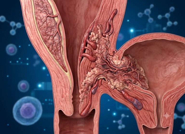
EMPD is a rare, slow-growing skin cancer (an intraepithelial adenocarcinoma) that arises in the skin’s surface layer, in the apocrine sweat glands. It mainly appears on the vulva, scrotum, perianal area, or occasionally the armpits.
Under the microscope, both EMPD and breast Paget’s disease contain “Paget cells,” large, pale malignant cells. But the breast form is almost always tied to an underlying ductal carcinoma, whereas EMPD is often a primary skin cancer, though sometimes a red flag for an internal cancer of the bladder, rectum, or prostate that has spread to the skin.
EMPD is frustrating because it looks like a benign condition: a red, scaly, or crusty plaque that may itch or burn. It is often misdiagnosed as eczema, psoriasis, or a stubborn fungal infection, delaying diagnosis by years. A biopsy is usually done only when the “rash” fails to respond to topical steroids or antifungal creams. Because EMPD can have “skip lesions” (cancer not visible to the naked eye), surgery often needs wide margins or specialized techniques such as Mohs surgery. While skeletal Paget’s affects mobility, EMPD calls for cancer surveillance to make sure it hasn’t become invasive or linked to an internal tumor.
4. Warmth Over a Bone Can Be an Early Sign
A distinctive early marker is localized warmth, or “hot spots,” on the skin over an affected bone. The hyperactive bone cells need a huge supply of oxygen and nutrients, so the body increases the number and size of blood vessels in the area (hypervascularity). If you feel unusual warmth over a bone, most often the shin or thigh, along with a deep, persistent ache, it may indicate active disease. It reflects the heart sending extra blood to the remodeling “construction zone.”
That extra demand can strain the heart. In rare, severe cases involving multiple bones, it can lead to high-output heart failure, where the heart is structurally healthy but can’t keep up with the disease’s demand for blood flow.
Noticing this warmth matters because it often appears before visible bowing or fractures, and monitoring it can help a physician judge whether the disease is active and needs bone-regulating medication such as bisphosphonates.
5. It Can Affect Hearing and Vision
When the skull is involved, the concern shifts from bone pain to protecting sensory function. The skull contains narrow channels that protect the cranial nerves, and as bone expands and thickens, these channels constrict.
Hearing loss. The most frequent neurological complication, and it happens two ways: the bone around the inner ear (the cochlea) can thicken and harden, interfering with sound conduction, and expanding bone can compress the auditory nerve. People often notice they’re turning up the TV or struggling to follow conversation in crowded rooms. Tinnitus (ringing or buzzing) and vertigo can accompany it.
Vision problems. Less common, but a medical priority. Bone growth around the optic foramen, the opening for the optic nerve, can compress the nerve and cause gradual vision loss, sometimes preceded by chronic, severe headaches from raised pressure inside the skull or increased blood flow to the bone.
Because these changes are slow, they’re often blamed on normal aging. A classic early clue is hats, helmets, or glasses becoming tight or no longer fitting. Combined with new sensory changes, this suggests active skull involvement and warrants imaging to assess the risk to your nerves.
6. The Cause Involves Both Genes and Possibly Viruses
Medicine hasn’t pinpointed a single origin. The prevailing theory is a “double hit” of genetic susceptibility plus an environmental trigger, which would explain why some people with similar backgrounds develop it and others don’t.
Genetics. Between 15% and 40% of patients have at least one first-degree relative with the disease. The most commonly identified marker is a mutation in the SQSTM1 gene, which produces a protein that helps regulate the “on/off” switch for osteoclasts. When it’s mutated, osteoclasts become hyper-responsive to growth signals, driving the rapid, disorganized bone destruction.
Viral trigger theory. Researchers have long suspected a slow-acting viral infection acts as the “spark” in genetically predisposed people. The leading theory involves the paramyxovirus family, which includes measles and respiratory syncytial virus (RSV). Viral-like particles have been found in the nuclei of affected osteoclasts, suggesting a virus may lie dormant in bone cells for decades. Interestingly, as measles vaccination has spread, the incidence and severity of Paget’s disease have declined in some regions, which is circumstantial evidence for the connection.
Age. The disease is rarely diagnosed before 50, suggesting that accumulated environmental exposure and a maturing immune system are needed for it to appear.
7. Prevention Focuses on Slowing Progression and Protecting Bone
Because the cause involves factors we can’t fully control, “prevention” mainly means slowing the remodeling disorder and protecting the surrounding skeleton. Since the affected bone is weaker, the aim is to build a “fortress” of healthy bone around it to prevent fractures and bowing.
- Calcium and vitamin D. These are the skeleton’s main building blocks, and demand rises when remodeling is overactive. Dairy, leafy greens, and fatty fish help. Because many adults are vitamin D deficient (and it’s needed to absorb calcium), a physician may recommend supplements.
- Weight-bearing exercise. Walking, hiking, or gentle resistance training stresses the skeleton in a healthy way, encouraging stronger bone and better muscle support for joints. For hip involvement, strong glutes and leg muscles help stabilize the joint and reduce strain on the misshapen pelvis.
- Routine monitoring. For people over 50 or with a family history, regular blood tests of alkaline phosphatase (ALP) can reveal the disease long before symptoms. If caught early, bisphosphonates can “shut down” the overactive cells and help prevent the disorganized bone structure from forming.
- Genetic counseling. Given the hereditary link, counseling can help you understand your risk and set up a personalized screening schedule, so the disease is managed proactively rather than after a “hot spot” or deep ache appears.
8. It’s Linked to Osteoporosis
Paget’s disease causes abnormal bone growth, but it also weakens bone density, so it can contribute to osteoporosis, a condition where bones become fragile and brittle. The imbalance between bone breakdown and formation produces bones that are larger but weaker, and the excessive turnover and irregular structure can lower density and raise fracture risk, even with minimal trauma.
The two share risk factors, including aging, family history, and hormonal changes (especially after menopause), and both are more common after 50. They’re different conditions, but they can occur together, which complicates bone health management.
How Paget’s contributes:
- Excessive turnover: the high rate of bone breakdown means new bone is weaker and less dense.
- Weakened structure: abnormal remodeling produces larger bones more prone to deformity and fracture.
- Increased fracture risk: fractures occur more easily, especially in the spine, pelvis, and legs.
Managing both: Bisphosphonates (such as alendronate) slow bone breakdown and help both conditions. Patients are also encouraged to eat a diet rich in calcium and vitamin D, do weight-bearing exercise, and avoid smoking and heavy drinking.
9. It Can’t Be Cured, but It Can Be Controlled
Strictly speaking, no treatment reverses bowing or restores hearing already lost to nerve compression. But bisphosphonates are highly effective at calming the remodeling process and can put the disease into long-term remission by normalizing the activity of the overactive bone cells. Most people live full, active lives provided they get early treatment to prevent further damage.
Conclusion
Understanding Paget’s disease matters for anyone affected or at risk. It doesn’t always cause obvious symptoms, but recognizing early signs such as bone pain, deformities, and joint stiffness can lead to earlier diagnosis and more effective treatment. With medications that regulate bone turnover and healthy lifestyle habits, many people manage their symptoms and stay active.
The exact cause remains unclear, but research continues to improve understanding and treatment. If you notice concerning symptoms or have a family history, see a healthcare provider for an accurate diagnosis and personalized care. Early intervention can prevent complications such as arthritis, fractures, and neurological problems.
Frequently Asked Questions
Can Paget’s disease be cured?
Not in the strict sense. Nothing reverses bone bowing or restores hearing already lost to nerve compression. But bisphosphonates are highly effective at calming the disease, often producing long-term remission by normalizing overactive bone cells. Most people live full, active lives with early treatment.
What does the blood test result mean?
The key marker is alkaline phosphatase (ALP), an enzyme released into the blood when bone turnover is high or the liver is stressed. If ALP is significantly elevated but liver function tests are normal, it strongly suggests the rapid bone breakdown and haphazard rebuilding typical of Paget’s disease.
Why is it called “pagetoid” disease?
“Pagetoid” is a descriptive term pathologists use for cells that look large, have clear halos, and appear to “spread” individually through tissue layers. Sir James Paget first described this pattern in the skin, which gave extramammary Paget’s disease its name. The term is now also used for the disorganized cellular behavior in both bone and skin conditions, even though they are unrelated.
Is Paget’s disease contagious?
No. Although the viral theory suggests a virus such as measles might act as a trigger, you can’t “catch” Paget’s from someone. It is a metabolic and genetic disorder that needs a specific predisposition, such as the SQSTM1 mutation, to appear. It is a slow-developing problem of the bone’s recycling system, not an active infection.
How often should I be screened if I have a family history?
Because of the strong hereditary component, proactive monitoring matters for relatives. If a parent or sibling has the condition, most specialists recommend starting screening around age 40, typically with an alkaline phosphatase blood test every two to three years. Catching the disease in its early “lytic” phase, before bone enlarges or bows, is the best way to prevent complications.