Cystoscopy: Types, Purpose, Procedure, Risks, and Recovery
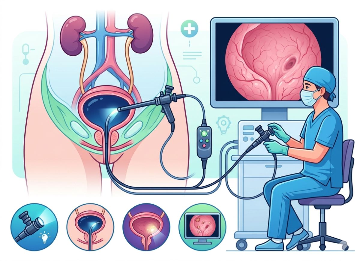
Blood in your urine, constant trips to the bathroom, or pelvic pain with no clear cause can be worrying. If you have any of these, your doctor may suggest a cystoscopy, a test that lets them look straight into your bladder and urethra to find out what’s going on.
The idea of a scope going into your urinary tract sounds scary, but cystoscopy is routine, minimally invasive, and gives doctors information other tests can’t.
Doctors use it to find bladder infections, stones, tumors, and structural problems. It can also treat: during the same procedure, a urologist can remove small growths or take tissue samples for testing. Roughly 1.5 million cystoscopies are done in the US every year.
There are two main types. The flexible scope is gentler and far more common, while the rigid scope is kept for more complex cases. The procedure usually takes only a few minutes, and most people go home the same day, though some mild discomfort or urinary symptoms can linger briefly.
Below, we cover the procedure, its risks, and recovery, so you know what to expect if your doctor recommends one.
What Is A Cystoscopy?
A cystoscopy is a test in which a urologist looks directly at the lining of your bladder and urethra, the tube that carries urine out of the body. The doctor uses a cystoscope, a slim instrument with a light and a lens or tiny video camera at its tip. It shows the inside of the urinary tract in real time, which X-rays and ultrasounds can’t do.
Cystoscopes come in two forms: a flexible, bendable tube and a straight, rigid tube that’s used when a biopsy or minor procedure is needed. The choice depends on what the doctor needs to do.
A urologist may recommend a cystoscopy to investigate, diagnose, monitor, or treat problems in the lower urinary tract. It is often used to find the cause of symptoms such as blood in the urine (hematuria), repeated urinary tract infections (UTIs), incontinence, overactive bladder, painful urination (dysuria), and long-term pelvic pain. Looking at the source directly can reveal inflammation, stones, or growths.
It is also key for diagnosing bladder and urethral conditions, including bladder stones, bladder cancer, non-cancerous polyps, cystitis (inflammation), bladder diverticula (small pouches in the bladder wall), and urethral strictures (narrowing of the urethra). People who have been treated for bladder cancer have regular cystoscopies too, so the urologist can catch any return of the disease early.
In some cases, cystoscopy is used for treatment. Through the scope, a urologist can remove small stones or tumors, take a biopsy, or inject medication into the bladder wall.
How Do You Prepare for a Cystoscopy?
Preparation is usually simple. Your urologist will tell you how much to drink, whether to adjust any medications, and that you’ll need to give a urine sample beforehand. Following these steps helps keep the procedure safe and as comfortable as possible.
You may be asked to drink some water before your appointment and then empty your bladder just before the test. This gives the team a clean sample to check for infection. If you have a UTI, the procedure may be postponed unless it’s an emergency. During the test, the doctor fills your bladder with sterile fluid to get a better view.
Tell your doctor about everything you take, including prescriptions, over-the-counter drugs, and supplements. You may need to pause blood thinners such as aspirin, clopidogrel (Plavix), or warfarin (Coumadin) for a few days beforehand, which lowers the risk of bleeding, especially if a biopsy is planned. Never stop a medication unless your doctor tells you to.
To help prevent infection afterward, your doctor may give you a single dose of antibiotic an hour or two before the procedure. This is especially common if you get frequent UTIs or have other risk factors. Take it exactly as instructed so it works properly.
Step-by-step During The Cystoscopy Procedure
You’ll lie on an examination table while the urologist gently passes the cystoscope through your urethra into your bladder, then uses sterile fluid to expand the bladder for a clear view. A diagnostic cystoscopy is quick, usually 5 to 15 minutes.
When you arrive, you’ll give a urine sample to check for infection and then change into a gown from the waist down. You’ll lie on your back with your knees bent and your feet in supports, which gives the urologist the best access to the urethra.
Next, a gel containing a local anesthetic such as lidocaine is applied at and inside the urethral opening. It numbs the area and also lubricates it so the scope slides in more easily. After a few minutes, the urologist gently guides the scope tip into the urethra and slowly moves it into the bladder. You may feel pressure or a strong urge to urinate at this point.
Once the scope is in the bladder, sterile water or saline flows through a channel in the scope and stretches the bladder wall so the whole lining is visible. You may feel full and want to urinate again. The urologist then checks the entire bladder surface for tumors, stones, inflammation, or ulcers while the camera sends images to a monitor. If something looks suspicious, tiny instruments can be passed through the scope to take a biopsy.
Is a Cystoscopy Painful?
Most people don’t find it painful. It can feel uncomfortable, with pressure and a strong urge to urinate, but the numbing gel keeps this manageable. Most patients say it’s more uncomfortable than painful. How it feels depends on your pain tolerance, how anxious you are, and whether a flexible or rigid scope is used.
Before the scope goes in, a generous amount of numbing gel is applied to the urethra, the most sensitive part of the urinary tract during this test. You may still feel the scope moving, but sharp pain is greatly reduced. Local anesthesia is enough for most outpatient diagnostic cystoscopies, while sedation or general anesthesia may be offered for a rigid cystoscopy or extensive biopsies.
Expect pressure and movement as the scope goes in, and a feeling of fullness with a strong urge to urinate as the bladder fills. Both are normal. Try to relax your pelvic muscles, because tensing up makes it more uncomfortable, and if you feel significant pain, tell your doctor or nurse right away.
The type of scope also matters. A flexible scope is thinner and bends with the curves of the urethra, so it’s usually more comfortable, especially for men. A rigid scope is wider and causes more pressure, but it’s needed for procedures like biopsies or stone removal because its channels are larger. If one is used, your doctor may discuss extra anesthesia.
Potential Complications and Side Effects of Cystoscopy
Side Effects After a Cystoscopy
Burning or pain when urinating is the most common complaint, caused by irritation of the urethral lining. It’s strongest the first few times you urinate and should ease within 24 to 48 hours. Drinking plenty of water dilutes your urine and makes it less painful.
A little blood in the urine, giving it a pink or light red color, is also very common, and you might see a few small clots. It comes from slight irritation of the urethra or bladder lining. After a biopsy, a bit more bleeding is expected. It usually clears in a day or two, but heavy bleeding or large clots are not normal and should be reported to your doctor.
Your bladder may also feel more sensitive, so you may need to go more often or more urgently than usual. This normally settles within a day, and staying hydrated helps flush the bladder.
More Serious but Rare Risks
Bacteria can enter the urinary tract during the procedure, but infection is uncommon, especially when preventive antibiotics are used. If one does develop, it can be more serious than an ordinary UTI and may spread to the kidneys (pyelonephritis) or, very rarely, the bloodstream (sepsis). Warning signs are fever, chills, persistent severe pain, and cloudy or foul-smelling urine.
In extremely rare cases, the scope can tear the wall of the urethra or bladder. The risk is slightly higher with a rigid scope or a complex procedure like tumor removal. A small tear may heal on its own with a temporary catheter, while a larger one may need surgical repair. Symptoms include severe abdominal pain, being unable to urinate, and fever.
Pink urine is normal, but significant bleeding is rare and mostly happens after a large biopsy or tumor removal. Bright red urine with many large clots may signal a serious bleed that needs medical treatment.
Some people, especially men with an enlarged prostate, can’t urinate for a while afterward because swelling and irritation block the flow. If you can’t pass any urine for several hours despite feeling the urge, call your doctor or go to the emergency room, since you may need a temporary catheter.
When Should You Call a Doctor After a Cystoscopy?
Contact a doctor right away if you develop a high fever with chills, can’t urinate at all, or see bright red blood or large clots in your urine. Mild discomfort is expected, but these symptoms need prompt attention. Your care team will give you a list of warning signs, so take it seriously.
A temperature above 101°F (about 38.3°C), especially with chills and body aches, is the main sign of infection. UTIs can escalate quickly, and early antibiotic treatment stops them from reaching the kidneys or bloodstream.
If you feel a strong, painful urge to go but can’t pass any urine for more than six to eight hours after the procedure, you may have urinary retention. The bladder needs to be drained promptly to avoid damage to the bladder or kidneys.
Some burning is normal, but severe or escalating pain in your abdomen, pelvis, or back is not. Pain that over-the-counter medication doesn’t relieve could point to a perforation or a serious infection. Likewise, pink urine is expected, but urine that stays bright red, or several large, thick clots, means significant bleeding that needs urgent assessment.
Cystoscopy Recovery: What to Expect After the Procedure?
Immediate Cystoscopy Recovery Period
Recovery starts quickly. Most people go home within 30 minutes to an hour. Because local anesthesia is most common, there’s no sedation to wear off, so long monitoring isn’t needed.
After the scope is removed, you’ll get privacy to dress. A nurse may watch for any immediate problems such as significant pain or heavy bleeding, and will go over aftercare instructions and warning signs with you. You may be asked to urinate before leaving to confirm you aren’t retaining urine. The first time may sting and the urine may look pink, which is normal.
The most important instruction is to drink lots of water for the rest of the day. About two to three liters flushes the bladder, dilutes urine to make it less painful, and lowers the chance of infection.
If you only had local anesthesia, you can usually drive yourself home. If you were sedated, arrange for someone else to drive you, since your coordination and judgment will be affected for several hours. You can eat and drink normally straight away.
Guidelines for Home Care and Activities
Recovery is usually fast, and a few simple steps make it easier. Your doctor will give you specific instructions, particularly if you had a biopsy or another procedure.
The most important step is to drink plenty of fluids. Aim for at least eight large glasses of water in the 24 hours after the procedure, which clears out remaining blood, reduces stinging, and lowers infection risk. To soothe the burning, try a warm bath or hold a warm, damp washcloth against the urethral opening. Acetaminophen (Tylenol) or ibuprofen (Advil, Motrin) can ease cramping or discomfort, but follow the package directions and check with your doctor first.
For the first 24 to 48 hours, skip strenuous activity, heavy lifting, and high-impact exercise so your body can heal and bleeding doesn’t increase. It’s also often recommended to wait a few days to a week before sexual activity so the urethra can heal. There are usually no dietary restrictions, but if your bladder is easily irritated, you may want to avoid caffeine, alcohol, and spicy or acidic foods for a day or two.
How Long Does It Typically Take to Feel Normal Again?
After a standard diagnostic cystoscopy, most people feel normal again within 24 to 48 hours. Burning and pink urine are strongest in the first day and gradually clear within two days. Timing varies from person to person and depends on whether a biopsy or other procedure was done.
The first 24 hours are when discomfort is most likely. Urinating may burn and your urine may be pink, and drinking plenty of water makes a real difference. Light activities like walking, desk work, and household chores are fine.
By day two, most people feel back to normal. Urination no longer hurts, urine has returned to its usual clear yellow, and frequency and urgency have faded. If symptoms aren’t improving or are getting worse after 48 hours, contact your doctor’s office. After a biopsy or tumor removal, these effects may last a bit longer, perhaps three to five days.
Men, especially those with an enlarged prostate, may have discomfort for slightly longer because the male urethra is longer. A prolonged or painful recovery isn’t typical, though, and lingering symptoms should be checked by your urologist.
When and How to Receive Your Cystoscopy Results?
You’ll often hear what the urologist saw right after the procedure. If a biopsy was taken, the full pathology results usually take several days to a week. How results are shared varies, so ask your urologist’s office about the process before you leave.
For many people, the visual inspection is the whole point. Once it’s over, the urologist can tell you whether the bladder lining looked healthy or whether they saw inflammation, stones, strictures, or suspicious growths. Hearing that nothing was found can be very reassuring. The urologist will discuss next steps in the recovery area, which might include more tests or a treatment plan.
If a biopsy was taken, the tissue goes to a lab, where a pathologist examines it under a microscope to see whether the cells are cancerous, pre-cancerous, or benign. This takes several business days, sometimes up to a week, so don’t assume that no news is good news.
Results are usually given at a follow-up appointment or by phone. A visit lets the doctor go through the pathology report, show you relevant images, and answer your questions. This matters most if the results point to a serious condition like bladder cancer, where the urologist will need to explain the diagnosis and outline a treatment plan. In other cases, a nurse or the doctor may phone you, so ask what their usual practice is.
Types of Cystoscopy
A Flexible vs. A Rigid Cystoscopy
The two types differ in how the scope is built and used, which affects your experience, the anesthesia needed, and the purpose of the procedure.
A flexible cystoscope is a thin, bendable fiber-optic tube that follows the natural curves of the urethra. It’s mainly used for diagnosis, such as finding the cause of blood in the urine, recurrent UTIs, or bladder pain. It’s usually done in an outpatient clinic while you’re awake, with only numbing gel. Because it bends, it’s much more comfortable, particularly for men, whose urethras curve naturally.
A rigid cystoscope is a straight, solid metal tube. It sounds more intimidating, but its wider channel lets larger, more complex instruments pass through. That makes it the preferred tool for treatment, such as biopsies, bladder stone removal, medication injections into the bladder wall, or removal of small bladder tumors. Because it can’t bend, it’s more invasive and is usually done in an operating room under regional anesthesia (like a spinal block) or general anesthesia, so you feel no pain. Its optics can also give a clearer, more magnified view of the bladder lining, which helps with detailed examination and precise surgical work.
The Cystoscopy For Men and Women
The procedure differs between men and women mainly because of the urethra.
The female urethra is short, about 1.5 to 2 inches, and runs straight from the bladder to the outside. This makes it simpler and quicker to pass either type of scope. A diagnostic flexible cystoscopy in women can often be finished in a few minutes with minimal discomfort, usually with just numbing gel. The direct route means less manipulation of the scope, which shortens the procedure and reduces irritation.
The male urethra is much longer, around 8 inches, and follows an S-shaped curve through the penis and prostate before reaching the bladder. That makes it a bigger technical challenge. Getting past the prostate and bladder neck takes more skill and can be more uncomfortable, so the flexible scope is strongly preferred for men’s diagnostic exams, since its tip can be steered around the curves. Even so, men may feel more pressure or urgency as the scope passes the prostate. When a rigid cystoscopy is needed, it’s almost always done under sedation or general anesthesia so the patient stays comfortable and still and the straight instrument can pass safely through the curved anatomy.
A Cystoscopy With A Biopsy or Stent Removal
Cystoscopy can do more than look. It can also be used for minor procedures like taking a biopsy or removing a ureteral stent.
If the urologist spots a polyp, suspicious growth, or inflamed patch on the bladder lining, they may take a biopsy. A long, thin instrument with tiny forceps is passed through the scope’s working channel to snip off a small piece of tissue, which goes to a lab to check for cancer cells, inflammation, or other disease. Biopsies are most often done with a rigid scope under regional or general anesthesia, since they can cause slight discomfort and minor bleeding. Afterward, pink or red-tinged urine for a day or two is common.
A ureteral stent is a thin, flexible plastic tube placed in the ureter to help drain urine from the kidney to the bladder, often after kidney stone surgery or to relieve a blockage. When it’s no longer needed, it can be removed quickly with a flexible cystoscope. After numbing gel is applied, the scope goes into the bladder and finds the lower end of the stent. A small grasper passes through the scope to hold the stent, and the scope, grasper, and stent are then gently withdrawn together. It’s very fast, often under a minute, and is usually well tolerated with only local anesthesia in the office.
Cystoscopy Compared to a Bladder Ultrasound or CT Scan
Cystoscopy, bladder ultrasound, and CT scans all help evaluate the urinary system, but they show different things. The key difference is direct viewing versus indirect imaging.
Cystoscopy gives the urologist a live, high-resolution video view inside the bladder. It’s considered the gold standard for examining the urothelium (the bladder’s inner lining) and the urethral opening. Its biggest strength is spotting subtle changes, such as flat, reddish patches that can indicate carcinoma in situ (an early-stage bladder cancer) and often don’t show on scans. It’s also the only one of the three that allows a biopsy for a definitive diagnosis. The downside is that it’s invasive, with a small risk of infection, bleeding, and discomfort.
A bladder ultrasound uses high-frequency sound waves to image the bladder. It’s good for checking the bladder’s shape and wall thickness, spotting large tumors or stones, and measuring how well the bladder empties (post-void residual urine). It’s safe, quick, and uses no radiation.
A CT scan, often a CT urogram, uses X-rays and computer processing to produce detailed cross-sections of the whole urinary tract, including the kidneys, ureters, and bladder. It’s better at locating bleeding that starts higher in the urinary system and at assessing structures outside the bladder.
Both ultrasound and CT are less sensitive than cystoscopy for small or flat tumors on the bladder lining. They show structure, but not the true color, texture, or blood supply of the tissue, and neither can take a biopsy.
FAQs
1. What is the main reason for cystoscopy?
Its main purpose is to examine the bladder and urethra so doctors can diagnose urinary problems. It’s commonly used to investigate unexplained symptoms such as blood in the urine (hematuria), frequent or painful urination, pelvic pain, or difficulty urinating.
It also helps identify and treat bladder stones, bladder cancer, infections, tumors, and abnormal growths. In some cases it’s used for treatment as well, such as removing stones, taking biopsies, or draining abscesses, so it serves both diagnostic and therapeutic roles.
2. Do they put you asleep for a cystoscopy?
Usually not. Most cystoscopies use local anesthesia, so you’re awake but shouldn’t feel pain in the bladder or urethra. If you’re anxious, a mild sedative can be given.
For more involved procedures, such as removing larger bladder stones or taking a biopsy, general anesthesia or heavier sedation may be used. Talk through your options with your provider beforehand so you can choose what suits you best.
3. Is cystoscopy a high-risk surgery?
No. It’s generally low-risk, especially as a diagnostic test. Complications are rare but can include infection, bleeding, injury to the urethra or bladder, and, very rarely, bladder perforation.
The risk goes up with more invasive uses, such as tumor removal or biopsy. Still, in the hands of a skilled urologist it’s usually safe, and complications can normally be managed quickly. The overall risk is far lower than for major surgeries.
4. What not to do after a cystoscopy?
Avoid anything that could strain the bladder or urethra. For the first 24 to 48 hours, skip heavy lifting, strenuous exercise, and sexual activity. Mild pain or burning when you urinate is normal and should gradually fade.
Stay well hydrated and avoid caffeine and alcohol, which can irritate the bladder. If you have severe pain, heavy bleeding, or signs of infection such as fever and chills, contact your healthcare provider right away.
5. How much does a cystoscopy cost?
Cost varies widely with location, whether it’s done in a clinic or hospital, and how complex the case is. In the US, a cystoscopy typically runs from about $1,000 to $3,000, and can be higher if extra treatment such as a biopsy or stone removal is involved.
Check with your insurer, since many plans cover it when it’s medically necessary. If you’re uninsured or underinsured, some providers offer payment plans or sliding-scale fees.
6. How many hours is a cystoscopy?
It doesn’t take hours. The diagnostic part usually lasts 10 to 30 minutes depending on the case, and a little longer if a biopsy or stone removal is added.
It’s normally an outpatient procedure, so you can go home the same day. Recovery is short, and most people are back to normal activities within a day or two, though mild discomfort may last a little while.
7. Is a CT scan better than a cystoscopy?
Neither is better overall, because they do different jobs. A CT scan produces detailed cross-sectional images and can detect bladder abnormalities, kidney stones, or other problems in the urinary tract, but it can’t show the inner lining of the bladder directly.
Cystoscopy gives a direct view of the bladder and urethra, which is essential for diagnosing conditions like bladder cancer, infections, or stones. Your doctor will choose based on your symptoms and medical history.
8. How do you stay calm during a cystoscopy?
Feeling anxious is natural. Start by sharing your worries with your doctor, who can walk you through each step and reassure you. Deep breathing or mindfulness techniques can also help you relax.
If you’re very nervous, ask about a mild sedative or extra local anesthetic. Remembering that the procedure is quick and low-risk can help too.
9. How many times can you have a cystoscopy?
There’s no fixed limit, especially when it’s medically necessary. People who need regular monitoring, such as those with recurrent bladder cancer, repeated UTIs, or stones, may have many cystoscopies over their lifetime.
Your provider will decide how often you need one based on your health, symptoms, and the risks of repeat procedures. Frequent cystoscopies should be weighed carefully, and alternatives may be considered where appropriate.
10. Should I be nervous about a cystoscopy?
It’s normal to feel nervous, but cystoscopy is usually low-risk and only mildly uncomfortable. It’s quick, and the discomfort is typically limited to pressure during the test and some burning when you urinate afterward.
If you have concerns, tell your provider, who can suggest ways to make you more comfortable. Most people find it easier than they expected, with a quick recovery and little downtime.
Conclusion
Cystoscopy is a valuable test that lets doctors look inside the bladder and urethra to diagnose and treat urinary tract conditions. It may sound intimidating, but it’s typically quick, low-risk, and minimally invasive, and it gives essential insight into your urinary health.
Knowing what happens before, during, and after the procedure helps you approach it with confidence, whether you’re investigating new symptoms or having a follow-up exam.
If you still have worries, talk to your healthcare provider. They can ease your concerns and make sure you’re fully prepared.