Intubation Made Simple: A Guide to the Breathing Tube Procedure
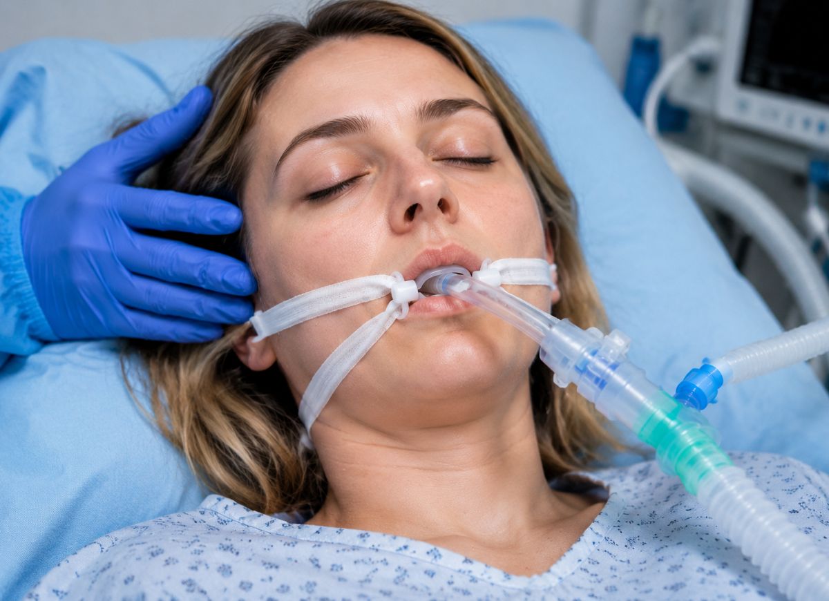
For many people, hearing the term breathing tube can be worrying or confusing. You may have encountered the word “intubation” in a hospital, during an emergency, or while watching a medical program. Intubation is a common procedure used when someone cannot breathe adequately on their own because of surgery, serious illness, or an emergency. Knowing what the procedure involves can make the experience easier to understand for patients and their loved ones.
Intubation is performed in hospitals around the world every day in emergency departments, operating rooms, and intensive care units. The procedure involves placing a thin tube into the windpipe, or trachea, so oxygen can reach the lungs and the airway can remain open. It may be necessary for people experiencing respiratory failure, severe breathing problems, or those receiving general anesthesia during surgery.
Although intubation may sound complicated, trained healthcare professionals perform it using specialized equipment and continuous monitoring. Depending on the situation, patients may receive sedation or anesthesia to reduce discomfort and awareness during placement. The breathing tube can remain in place for several hours or multiple days depending on the patient’s condition, while the medical team monitors oxygen levels, vital signs, and lung function.
This guide explains intubation in straightforward language, including why it is performed, how the procedure works, what it may feel like, possible risks, recovery, and alternatives. Understanding these steps can make the process less frightening and help patients and caregivers know what to expect during critical medical care.
What are Reasons for Intubation?
Intubation is used to secure and protect the airway when a person cannot breathe safely or effectively without assistance. The three main reasons include supporting patients with respiratory failure, protecting the airway from blockage or aspiration, and controlling breathing during surgery under general anesthesia.
The need for intubation can arise from many different medical conditions. Understanding these situations helps explain why a breathing tube may be necessary and whether the procedure is being performed as part of a planned treatment or an emergency response.
Common Medical Conditions That Require Intubation
Medical conditions that require intubation generally fall into three broad groups: respiratory failure, airway protection, and planned anesthesia. These categories help doctors identify the main reason a patient needs an artificial airway.
Respiratory failure is one of the most common reasons for intubation in critical care. It occurs when the lungs are unable to exchange gases properly, meaning the body is not receiving enough oxygen or is unable to remove enough carbon dioxide. Several serious illnesses can lead to this condition.
Acute Respiratory Distress Syndrome (ARDS) is a severe form of lung injury involving widespread inflammation. Fluid can collect in the tiny air sacs of the lungs, making breathing difficult and reducing oxygen exchange. Intubation and mechanical ventilation can provide breathing support while doctors treat the underlying cause, such as severe pneumonia or sepsis.
Severe pneumonia can also lead to respiratory failure. The infection may cause the lung air sacs to become inflamed and fill with fluid or pus. When the illness becomes severe, a patient may become too weak to breathe effectively or maintain safe oxygen levels, making mechanical ventilation necessary.
During a serious Chronic Obstructive Pulmonary Disease (COPD) exacerbation, breathing can become extremely difficult and carbon dioxide may build up in the bloodstream. This can cause respiratory acidosis and may require intubation to reduce the workload on exhausted breathing muscles and restore safer gas exchange.
Airway protection is another important reason for intubation. A patient may be unable to keep the airway open or may be at risk of inhaling stomach contents, saliva, or other material into the lungs.
People who are unconscious because of a head injury, stroke, overdose, or another medical condition may lose protective reflexes such as coughing and gagging. A breathing tube can help keep the airway open and reduce the risk of aspiration.
Severe injuries involving the face, neck, or chest may also cause bleeding, swelling, or structural damage that threatens the airway. In these situations, clinicians may place a breathing tube before swelling or obstruction becomes more severe.
General anesthesia during major surgery is another common reason for planned intubation. The medications used during general anesthesia produce unconsciousness and can temporarily relax or paralyze the muscles responsible for breathing. A breathing tube allows the anesthesiologist and ventilator to control oxygen, carbon dioxide, and breathing throughout the operation.
Is the Intubation Procedure Always an Emergency?
Intubation is not always performed during an emergency. It can be planned ahead of time as an elective procedure or carried out urgently when a patient suddenly develops life-threatening breathing or airway problems. The circumstances determine how quickly the team must act and how the procedure is prepared.
Elective intubation is usually scheduled in advance, most commonly for major surgery requiring general anesthesia. It generally takes place in an operating room where the healthcare team has enough time to evaluate the patient’s airway, review medical history, prepare equipment, and anticipate possible difficulties.
The patient is connected to monitoring equipment before medications are given. The process is carefully organized, and the necessary airway equipment and trained professionals are immediately available. The goal is to establish a secure airway throughout the operation while keeping the patient stable.
Emergency intubation, by comparison, is performed when immediate airway or breathing support is needed. It may be necessary during cardiac arrest, severe respiratory failure, major trauma, or a serious allergic reaction such as anaphylaxis. Depending on the circumstances, it can take place in an emergency department, intensive care unit, or even before the patient reaches the hospital.
Because emergency situations are time-sensitive, clinicians may have very little information about the patient’s medical history or airway. Rapid Sequence Intubation (RSI) is commonly used, involving medications that quickly produce unconsciousness and muscle relaxation so the airway can be secured rapidly. The immediate priority is restoring adequate oxygenation and ventilation.
The Intubation Process
Intubation follows a carefully coordinated series of steps. The medical team first prepares the patient with oxygen and appropriate medications, positions the patient, visualizes the airway using specialized equipment, places the breathing tube into the trachea, and then confirms that the tube is correctly positioned.
From the patient’s perspective, the process is usually not consciously experienced because sedation or anesthesia is generally given before the tube is inserted. Breaking the procedure into stages can make it easier to understand what happens during these critical moments.
Doctors carefully prepare patients before intubation to improve safety. This preparation may include pre-oxygenation, sedative and paralytic medications, and positioning the head and neck correctly.
This preparation is important because it helps reduce complications such as dangerously low oxygen levels or low blood pressure. Each step is performed with the goal of creating the safest possible conditions for placing the tube quickly.
Before medications are administered, the patient is usually given a high concentration of oxygen through a mask or another oxygen-delivery device. This increases the amount of oxygen stored in the lungs and creates a temporary reserve.
That oxygen reserve provides additional time for the clinician to place the tube before oxygen levels begin to fall during the brief period when the patient is not breathing independently.
Sedative and paralytic medications are commonly used during Rapid Sequence Intubation (RSI). An induction medication such as propofol, etomidate, or ketamine may be administered through an intravenous line to make the patient unconscious and unaware of the procedure.
A neuromuscular blocking medication, such as succinylcholine or rocuronium, may then be given. This temporarily relaxes the skeletal muscles, including those involved in the jaw and airway. Muscle relaxation helps prevent coughing or gagging and allows the clinician to visualize the vocal cords and place the tube more safely.
Positioning is another important part of the procedure. The patient’s head and neck are placed in a position that helps create the best possible view of the airway. Depending on the patient’s anatomy and medical condition, clinicians may use a slightly extended or specially supported position.
Proper alignment can improve visualization of the vocal cords during laryngoscopy and may make tube placement more efficient.
The Feeling of Intubation
Because sedation or anesthesia is commonly administered before intubation, most patients do not consciously feel the actual placement of the breathing tube. The medications are intended to produce unconsciousness, reduce discomfort, and prevent awareness of the procedure.
The medical team makes sure the appropriate medications have taken effect before attempting tube placement. These medicines work quickly and are selected according to the patient’s condition and the circumstances.
Although the insertion itself is generally not felt when adequate sedation is used, patients who remain intubated in an intensive care unit may notice the tube after sedation is reduced. Waking up while the tube is still in place can feel unusual or uncomfortable.
One of the most noticeable sensations may be pressure or irritation in the back of the throat. The tube can create an urge to cough or gag because the body naturally tries to remove foreign objects from the airway.
The endotracheal tube passes through the vocal cords, which means the patient cannot normally speak while it remains in place. This can be frustrating, particularly for patients who are awake and want to communicate with their family or healthcare team. They may need to use gestures, writing, or other communication methods.
The mouth and throat can also become dry because the patient may have the mouth slightly open while breathing is supported by a ventilator. Regular oral care from nursing staff helps reduce dryness and discomfort.
Patients who are confused or heavily sedated may sometimes have soft wrist restraints. These may be used as a safety measure to prevent accidental removal of the breathing tube, which can be dangerous.
Throughout mechanical ventilation, healthcare professionals monitor comfort and may use sedative and pain-relieving medications when appropriate to reduce anxiety, discomfort, and distress.
Immediate Risks During The Intubation Procedure
The immediate risks of intubation can include injury to the airway, incorrect placement of the breathing tube, and temporary changes in blood pressure or oxygen levels.
These complications can develop around the time of tube placement and require rapid recognition and treatment. Although medical teams use careful techniques and monitoring to reduce these risks, they cannot always be completely eliminated.
The laryngoscope and breathing tube can sometimes cause injury to the lips, tongue, throat, or other airway structures. Dental injury, including chipped or loosened teeth, can also occur, especially in people with existing dental problems. In rare situations, the vocal cords may become irritated or injured, which can result in temporary or, less commonly, lasting hoarseness.
Esophageal intubation is a serious complication in which the tube accidentally enters the esophagus instead of the trachea. If this is not recognized, air from the ventilator will enter the stomach rather than the lungs, preventing adequate oxygenation.
To reduce this risk, clinicians confirm tube placement immediately after insertion. They may use end-tidal carbon dioxide monitoring, listen to breath sounds on both sides of the chest, and check for symmetrical chest movement.
The medications and airway stimulation involved in intubation can also produce temporary physiological changes. Sedative medications may lower blood pressure, particularly in patients who are already seriously ill or dehydrated. The short period of apnea can also cause oxygen levels to fall, although pre-oxygenation helps reduce this risk.
Stimulation of the airway can sometimes affect heart rate and rhythm, potentially causing bradycardia or tachycardia. The medical team continuously monitors the patient and can provide fluids, medications, or other interventions when needed.
Long-term Complications of Intubation
Long-term complications are more closely associated with prolonged mechanical ventilation than with the brief act of inserting the breathing tube. Potential problems include ventilator-associated pneumonia, injury to the vocal cords or trachea, and weakening of the respiratory muscles.
The longer a breathing tube remains in place, the greater the potential for certain complications. The tube bypasses some of the body’s normal airway defenses and remains in contact with delicate tissues.
Ventilator-Associated Pneumonia (VAP) is an important potential complication of prolonged ventilation. Because the breathing tube bypasses natural defenses such as the nose and normal cough reflex, bacteria can have a more direct route into the lower respiratory tract.
Secretions can collect around the tube and may enter the lungs. Hospitals use several measures to lower the risk of VAP, including keeping the head of the bed elevated, providing regular oral care, and reducing ventilator use as soon as it is safely possible.
Vocal Cord and Tracheal Injury: The tube and its cuff can place ongoing pressure on the delicate tissues of the airway. Over time, this may cause irritation, inflammation, ulceration, or other forms of tissue damage.
Pressure-related injury can sometimes result in vocal cord problems, leading to a weak, breathy, or hoarse voice after extubation. Many cases improve, although more serious injuries may persist.
In uncommon cases, prolonged pressure from the tube cuff can injure the trachea and cause scar tissue. This may result in tracheal stenosis, a narrowing of the windpipe that can create breathing difficulties after the tube has been removed.
Mechanical ventilation can also reduce the amount of work performed by the patient’s own breathing muscles. When the diaphragm remains inactive for an extended period, it can become weaker.
This condition, known as ventilator-induced diaphragmatic dysfunction, can make it harder to discontinue mechanical ventilation. Physical and respiratory therapy may be used to help maintain strength and gradually encourage independent breathing when appropriate.
Recovery after Intubation
Removing a breathing tube is called extubation. It is a carefully monitored process in which the healthcare team first determines whether the patient can breathe independently and safely. A spontaneous breathing trial may be performed before the tube is removed.
Extubation is an important recovery milestone because it means the underlying problem has improved enough for the patient to begin breathing without the same level of mechanical support. The medical team continues to monitor the patient closely after removal to make sure breathing remains stable.
When It Is Safe to Remove The Breathing Tube?
Doctors consider several factors before deciding that a breathing tube can be safely removed. They evaluate whether the original medical problem has improved, whether the patient can breathe adequately without major ventilator support, and whether they can protect their own airway.
The weaning process involves gradually reducing ventilator support and assessing how well the patient responds. The final decision is based on the overall clinical picture rather than one measurement alone.
The original reason for intubation should have improved substantially or resolved. For example, a patient who was intubated because of severe pneumonia should show clinical evidence that the infection is improving and lung function is recovering.
The patient should also be able to maintain adequate oxygen levels with relatively little ventilator support. Oxygen saturation, oxygen requirements, and carbon dioxide levels may all be assessed to determine whether the lungs are functioning well enough without extensive mechanical assistance.
Cardiovascular stability is also important. The patient’s blood pressure and heart rate should be reasonably stable, and they should not require high doses of medications to support circulation.
The patient should generally be awake enough to follow commands and protect the airway. A strong cough and adequate ability to clear secretions are important because they help reduce the risk of aspiration after extubation.
Successful Spontaneous Breathing Trial (SBT) is an important final assessment. During an SBT, ventilator support is significantly reduced while the patient breathes largely on their own. The trial may last from about 30 minutes to a couple of hours depending on the patient’s condition and clinical protocol.
The medical team watches for signs that the patient is struggling, such as rapid breathing, falling oxygen levels, unstable heart rate or blood pressure, or visible respiratory distress. If the patient tolerates the trial successfully, the team may determine that extubation is appropriate.
Common Side Effects After the Breathing Tube is Removed
After extubation, the most common temporary effects include a sore throat, hoarse voice, and some difficulty swallowing. These symptoms are usually related to irritation and inflammation caused by the breathing tube.
The endotracheal tube can irritate the throat and larynx even when it has been used for a relatively short period. Healthcare professionals monitor these symptoms and provide supportive care as needed.
Sore throat (Post-Extubation Pharyngitis) is one of the most frequent complaints following extubation. The tube can irritate the sensitive tissues of the throat, producing a scratchy or raw sensation. Symptoms are often most noticeable during the first day or two and usually improve as the tissues recover. If swallowing is safe, cool liquids, ice chips, or appropriate throat remedies may provide relief.
A weak, raspy, or hoarse voice is also common immediately after the tube is removed. Because the tube passes between the vocal cords, temporary swelling and irritation can interfere with normal vocal cord movement. In many patients, the voice gradually returns to normal over several days.
Patients may be advised to limit excessive talking while their throat heals. If significant hoarseness continues for several weeks, an evaluation by an otolaryngologist (ENT specialist) may be appropriate.
Some people also have difficulty swallowing after extubation. The tube can temporarily reduce throat sensation, and prolonged intubation may weaken the muscles involved in swallowing. This can increase the risk of coughing or choking, particularly when drinking thin liquids.
Because aspiration can be dangerous, healthcare staff may perform a bedside swallow screening before allowing the patient to eat or drink. If problems are identified, a speech-language pathologist (SLP) may perform a more detailed assessment and recommend swallowing techniques or a modified diet until function improves.
Advanced Topics and Alternatives in Airway Management
Airway management includes more than standard oral intubation. Depending on the patient’s condition and how long breathing support is expected to be needed, doctors may consider different types of tubes, surgical airways such as tracheostomy, or non-invasive breathing support.
Understanding these options can help patients and families see why one method may be selected instead of another. The choice depends on the severity of illness, expected duration of respiratory support, airway anatomy, and other medical factors.
Communication When Patients Have a Breathing Tube?
An endotracheal tube passes through the vocal cords before entering the trachea. Because the tube occupies the space between the vocal cords, normal vocal cord movement is prevented.
For this reason, a patient generally cannot speak while the tube remains in place, even if they are awake and alert. This can be frustrating and may create anxiety or feelings of helplessness. ICU teams therefore use alternative communication methods to help patients express their needs.
Patients who are able to use their hands may communicate through a small whiteboard, marker, pen, or paper. If writing is difficult, picture or letter boards can allow patients to point toward words, objects, or needs.
Simple gestures can also be useful. Nodding, shaking the head, or using thumbs-up and thumbs-down gestures can help answer yes-or-no questions. Family members and healthcare workers may also interpret lip movements when the patient mouths words.
Intubation vs. Tracheostomy
Both intubation and tracheostomy can provide an airway for mechanical ventilation, but they differ in how they are performed and how long they are generally used.
Intubation is a non-surgical procedure in which a tube is inserted through the mouth or nose and passed into the windpipe. It is commonly used when an airway needs to be secured quickly, including during emergencies, surgery, or short-term critical care.
When an endotracheal tube is needed for an extended period, it can cause discomfort and irritation to the mouth, throat, and vocal cords. A tracheostomy involves surgically creating an opening in the front of the neck directly into the trachea. A specialized tube is then placed through this opening.
The decision to perform a tracheostomy depends largely on how long respiratory support is expected to be necessary and the patient’s overall condition.
A tracheostomy can be more comfortable for some awake patients and may reduce the amount of sedation needed. It also leaves the mouth available for oral care, and certain specialized tracheostomy tubes can allow speech when appropriate.
For some patients who need prolonged ventilator support, a tracheostomy can also make the process of weaning from the ventilator easier and allow a more gradual transition toward independent breathing.
Endotracheal vs. Nasotracheal Intubation
The main difference between endotracheal and nasotracheal intubation is the route used to place the breathing tube into the trachea. Endotracheal intubation, often called orotracheal intubation, involves passing the tube through the mouth.
Oral intubation is commonly preferred because it can usually be performed quickly and allows the use of a relatively larger tube. It is widely used during emergencies, general anesthesia, and critical care. Potential disadvantages include irritation of oral structures, dental injury, and discomfort.
Nasotracheal intubation uses a smaller, flexible tube that enters through the nose and travels into the trachea. This method may be selected for certain procedures when access to the mouth needs to remain unobstructed.
It can be particularly useful during some dental, oral, or maxillofacial procedures. It may also be considered when mouth opening is severely restricted because of trauma or conditions such as trismus.
In selected patients who need an airway for several days but are not candidates for tracheostomy, the nasal route may provide another option depending on the clinical situation.
BiPAP or CPAP Machine as An Alternative to Intubation
BiPAP or CPAP may sometimes provide an alternative to intubation for certain patients with breathing difficulties. These machines provide non-invasive ventilation (NIV), supporting breathing without placing a tube into the trachea.
CPAP (Continuous Positive Airway Pressure) and BiPAP (Bilevel Positive Airway Pressure) deliver pressurized air through a mask covering the nose, mouth, or both. The pressure can help keep the airways open, improve oxygenation, and reduce the effort required to breathe.
NIV is often considered when a patient is awake, cooperative, and able to protect their own airway and clear secretions safely.
However, non-invasive ventilation is not suitable for every patient with respiratory failure. If the illness is too severe or NIV does not provide enough support, invasive ventilation through intubation may become necessary.
Patients with severe conditions such as significant ARDS or profound shock may require invasive mechanical ventilation when non-invasive support cannot adequately improve oxygen or carbon dioxide levels.
FAQs
1. How serious is it to be intubated?
Being intubated is a significant medical intervention and usually means that a patient needs help maintaining adequate breathing or protecting their airway. It may be used during major surgery, emergencies, or serious critical illness and can be life-saving.
Although trained professionals perform intubation routinely, the procedure carries potential risks such as airway injury, infection, and complications associated with mechanical ventilation. Patients who are intubated require close monitoring of breathing, oxygen levels, vital signs, and airway function.
2. What is the purpose of intubation?
The main purpose of intubation is to secure the airway and help ensure that oxygen reaches the lungs. It allows healthcare professionals to control or support breathing when a patient cannot breathe effectively because of respiratory failure, trauma, anesthesia, or severe illness.
In this way, intubation provides temporary breathing support while doctors address the underlying medical problem. Depending on the patient’s condition, the tube may remain in place for a short time or for a longer period.
3. How long can someone stay intubated?
The length of time a patient remains intubated varies considerably. Some people need a breathing tube only for a few hours during surgery, while critically ill patients may require mechanical ventilation for days or longer.
As the duration increases, the risk of problems such as throat irritation, vocal cord injury, and infection can also increase. Healthcare teams regularly reassess whether the patient is ready to breathe without the tube.
4. Is being intubated painful?
Most patients receive sedation or anesthesia before intubation, so they generally do not experience pain during tube placement. Depending on the situation, especially during certain emergency or awake procedures, some pressure or discomfort may still be experienced.
After the tube is removed, temporary symptoms such as a sore throat, hoarseness, or throat irritation are common. These usually improve over several days as the airway heals.
5. Are people awake when intubated?
Most patients are sedated or under anesthesia during intubation. This helps reduce discomfort, anxiety, gagging, and movement while the tube is being inserted.
Awake intubation may be used in selected situations, particularly when doctors need to preserve spontaneous breathing because of a difficult or potentially complicated airway. The approach depends on the patient’s condition and the clinical circumstances.
6. What happens if intubation fails?
If standard intubation is unsuccessful, healthcare professionals have alternative methods for maintaining oxygenation and securing the airway. These may include bag-mask ventilation, different airway devices, or emergency surgical airway procedures such as cricothyrotomy.
Because failed intubation can become dangerous quickly, trained medical teams prepare backup strategies and respond rapidly when the first approach is unsuccessful.
7. Do they sedate you before intubation?
Yes, sedation or anesthesia is generally given before intubation to reduce awareness, discomfort, and the gag reflex. Medications such as propofol, etomidate, or other agents may be selected according to the patient’s condition and the urgency of the situation.
In unusual emergencies, medication choices may need to be adjusted depending on the patient’s condition and how quickly the airway must be secured. The goal is to establish the airway as safely and comfortably as circumstances allow.
8. What is the survival rate for intubation?
The outcome of intubation depends largely on the medical condition that made the procedure necessary rather than the tube itself. People who are intubated temporarily during surgery may have very different outcomes from patients who require ventilation because of severe critical illness.
Factors such as the underlying disease, overall health, organ function, and severity of illness all influence recovery. Close monitoring and timely treatment are important parts of care for intubated patients.
9. How long will my throat hurt after intubation?
A sore or scratchy throat is common after extubation. Some people also experience hoarseness, coughing, or mild irritation for several days, and symptoms may occasionally last longer depending on how long the tube was in place.
Drinking suitable fluids, resting the voice, and following the healthcare team’s advice may help with recovery. Persistent, severe, or worsening symptoms should be discussed with a healthcare professional.
10. What to do immediately after intubation?
After intubation, the medical team closely monitors oxygen levels, vital signs, breathing, and lung function. Once the patient is eventually extubated, recovery generally focuses on rest, hydration, airway monitoring, and gradual return to eating, speaking, and normal activity as appropriate.
Patients should follow their healthcare team’s instructions and report persistent breathing problems, significant throat pain, swallowing difficulties, or other unusual symptoms.
Conclusion
Intubation is an important life-saving procedure used to maintain breathing when a patient cannot breathe safely or effectively without assistance. Although the idea of a breathing tube can be intimidating, understanding why it is used, how it is placed, what patients may experience, and how recovery works can make the process easier to understand.
Most patients receive careful monitoring and appropriate sedation or anesthesia during the procedure. Temporary effects such as throat irritation or hoarseness are common after the tube is removed and often improve as the airway heals. By understanding the potential risks, recovery process, and available airway-support options, patients and caregivers can better navigate the period of critical care and recovery.