What Is Precancerous? 7 Things to Know About Abnormal Cell Changes
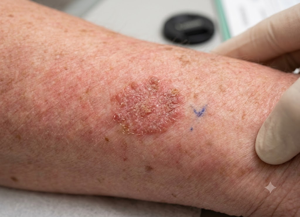
What is precancerous? The word generally refers to abnormal changes in cells that are not cancer but may have the potential to develop into cancer over time. These changes can be discovered through screening tests, biopsies, Pap tests, colonoscopies, skin examinations, or other medical evaluations. Although hearing the term can be concerning, precancerous does not mean that someone has cancer.
Learning about precancerous changes can make it easier to understand a diagnosis and follow the recommended care plan. Some abnormal cells may return to normal without treatment, while others can remain unchanged or require treatment and regular monitoring.
The level of concern depends on several factors, including where the abnormal cells are located, how they appear under a microscope, and whether risk factors such as HPV infection, smoking, chronic inflammation, or family history are present. Here are seven important things to know about abnormal cell changes and what a precancerous result can mean.
What are precancerous conditions?
A precancerous condition, sometimes called a pre-malignant condition, describes abnormal cell growth that has a greater-than-normal potential to become cancerous. It can be viewed as a stage between healthy tissue and invasive cancer, which gives doctors an opportunity to monitor or treat the abnormal cells before cancer develops.
To better understand what is precancerous, it helps to know the medical terms involved, how precancerous cells differ from cancer cells, and what can happen after an abnormality is discovered.
Pathological Transition: Defining the Pre-Malignant State
To understand what is precancerous, think of it as a possible step between healthy tissue and invasive cancer. In this state, cells have developed abnormal features that may increase their chance of becoming cancerous over time. These changes are often discovered during routine screening, such as a Pap test or colonoscopy, even when the person has no symptoms.
[Normal Epithelium] ──► [Dysplasia / Precancerous State] ──► [Invasive Carcinoma]
                                 │
                     (Confined by Basement Membrane)
One important term used when discussing what is precancerous tissue is dysplasia. Dysplasia describes abnormal cell development in which cells may differ from normal cells in their size, shape, organization, and maturity. Pathologists examine these changes under a microscope and may assign a grade based on how abnormal and disorganized the tissue appears.
For example, a low grade squamous intraepithelial lesion is a mild form of abnormal cell change that can occur in cervical tissue. With a low grade squamous intraepithelial lesion, the cells have some structural abnormalities but still maintain much of their normal organization.
If abnormal changes become more pronounced, they may develop into high-grade dysplasia. At this stage, the cells can look considerably different from normal cells and may show substantial disorganization. However, even severe dysplasia remains limited to the tissue layer where it developed and has not crossed the basement membrane.
Anatomical Boundaries: Precancerous vs. Cancerous Cells
One of the main differences between precancerous and cancerous cells is their ability to invade surrounding tissue and spread to other parts of the body. Both can have abnormal appearances and increased growth, but invasive cancer has crossed normal tissue boundaries.
                    [Anatomical Barrier Boundaries]
                                   │
     ┌─────────────────────────────┴─────────────────────────────┐
     ▼                                                           ▼
[Precancerous Cells (Dysplasia)]                    [Cancerous Cells (Malignancy)]
 ├── Abnormal growth                                  ├── Uncontrolled growth
 ├── Irregular cellular appearance                    ├── Often poorly differentiated
 └── Confined to the original tissue layer            └── Can invade surrounding tissue
Precancerous cells may grow and organize differently from normal cells, but they remain confined to their original tissue layer. They have not invaded deeper tissues or gained the ability to spread throughout the body.
Cancer cells, on the other hand, can invade surrounding tissues. Once cancer breaks through normal tissue barriers, it may gain access to blood vessels or lymphatic channels. Cancer cells can then travel to other parts of the body and form additional tumors, a process known as metastasis.
Precancerous cells can still retain some characteristics of the tissue in which they developed. Cancer cells may become increasingly abnormal and lose many of the features that identify their original tissue.
Prognosis and the Spectrum of Progression
A precancerous diagnosis does not mean that cancer will definitely develop. Instead, it identifies an abnormal area that may have a greater risk of progressing and gives healthcare professionals an opportunity to monitor or treat it.
The course of a precancerous change can vary. Some abnormalities may remain stable for years, some may improve or disappear, and others may progress toward cancer. The likelihood of progression depends on factors such as the location of the abnormal cells, their grade, the underlying cause, and individual risk factors.
Many mild abnormalities can resolve without treatment. For example, some low-grade cervical cell changes may disappear as the immune system clears an underlying HPV infection.
Because some mild changes improve naturally, doctors may recommend active surveillance, sometimes called watchful waiting. This means having follow-up examinations or tests at recommended intervals to make sure the abnormality is improving or not becoming more severe.
When high-grade abnormalities have a greater risk of progressing, treatment may be recommended. Depending on the location, doctors may remove or destroy the abnormal tissue. For example, a polypectomy can remove certain colon polyps during a colonoscopy, while a loop electrosurgical excision procedure (LEEP) can remove abnormal cervical tissue.
What are the critical facts about abnormal cell changes?
There are seven important facts that can help explain abnormal cell changes, including their causes, common types, symptoms, diagnosis, potential to reverse, treatment, and prevention. Understanding these areas can make the term precancerous less confusing and help people understand why follow-up care may be recommended.
Next, we will look at these seven areas in more detail to explain precancerous conditions and how they are managed.
Etiological Triggers: What Drives Cellular Mutation?
To understand what is precancerous tissue, it is useful to consider what causes cells to become abnormal. Environmental exposures, infections, inherited factors, and long-term inflammation can contribute to DNA damage. When genetic changes accumulate, normal controls over cell growth may become disrupted, which can contribute to dysplasia.
[Carcinogens / Chronic Inflammation / Persistent Viruses]
                          │
                          ▼
             [Accumulated DNA Copy Errors]
                          │
                          ▼
       [Changes in Growth-Control Genes]
                          │
                          ▼
    [Dysplasia / Precancerous Proliferation Stage]
Pathological Triggers of Abnormal Growth
Persistent Pathogenic Infections: Certain infections can contribute to abnormal cell changes. High-risk types of human papillomavirus (HPV), for example, can interfere with normal cell-growth controls and contribute to cervical dysplasia. Long-term infection with Helicobacter pylori can also cause chronic inflammation in the stomach and increase the risk of abnormal gastric changes.
Chronic Tissue Inflammation: Ongoing inflammation can repeatedly damage tissue and require cells to continually repair and replace themselves. Over time, this may increase the chance of abnormal cellular changes. Chronic acid reflux, for example, can damage the esophageal lining and contribute to Barrett’s esophagus, a condition associated with an increased risk of esophageal cancer.
Direct Carcinogen Exposure: Some substances and environmental exposures can damage cellular DNA. Tobacco smoke contains chemicals that can contribute to abnormal changes in tissues of the mouth and throat. Long-term exposure to ultraviolet (UV) radiation can also damage skin cells and contribute to precancerous growths such as actinic keratoses.
Pathological Mapping: Common Precancerous Manifestations
Precancerous changes can develop in different parts of the body. Many are identified through screening, routine examinations, or tissue testing.
                      [Anatomical Precancerous Grid]
                                     │
     ┌───────────────────────────────┼───────────────────────────────┐
     ▼                               ▼                               ▼
[Actinic Keratosis (Dermal)]  [Adenomatous Polyps (GI)]    [Cervical Intraepithelial (Gyn)]
 ├── Linked to UV exposure      ├── Develop in colon lining  ├── Often linked to high-risk HPV
 ├── Rough, scaly skin patch    ├── May become cancerous     ├── CIN 1 to CIN 3 grading
 └── Can precede skin cancer    └── Found during colonoscopy └── Evaluated with cervical testing
Actinic Keratosis (AK)
Actinic keratoses are rough, scaly patches that commonly appear on skin exposed to the sun, including the face, scalp, ears, and hands. A single lesion has a relatively low chance of becoming squamous cell carcinoma, but people who develop multiple lesions may have a higher overall risk of skin cancer.
Adenomatous Polyps (Colorectal Adenomas)
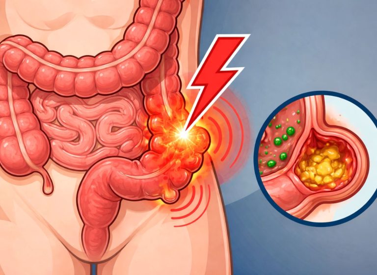
Adenomatous polyps are growths that develop in the lining of the colon or rectum. Some can gradually develop into colorectal cancer, which is why they are commonly removed when found during a colonoscopy. Removing certain precancerous polyps can help prevent them from progressing.
Cervical Intraepithelial Neoplasia (CIN)
Cervical intraepithelial neoplasia (CIN) describes abnormal cell changes on the surface of the cervix. These changes are often associated with persistent infection from high-risk HPV types. CIN is generally graded from CIN 1, which represents mild abnormalities, to CIN 3, which represents severe dysplasia.
Clinical Detection and the Silent Nature of Dysplasia
Many precancerous changes do not cause noticeable symptoms. Because abnormal cells can remain confined to their original tissue layer, a person may feel completely well even when significant cellular changes are present. This is one reason routine screening can be important for detecting abnormalities early.
[Suspicious Tissue Finding] ──► Tissue Biopsy ──► Histopathological Diagnosis
A diagnosis may involve several steps:
Initial Preventive Screening: Screening tests are designed to identify abnormalities before symptoms appear. Depending on the part of the body being evaluated, this can include cervical screening with Pap or HPV tests, colonoscopy for colorectal abnormalities, or mammography for breast changes.
Physical and Endoscopic Exams: Some abnormalities can be identified during a physical examination. For example, a dermatologist may recognize actinic keratoses during a skin examination. Endoscopic procedures can allow doctors to examine the lining of areas such as the esophagus, stomach, or colon.
The Definitive Tissue Biopsy: A biopsy involves removing a sample of tissue so that a pathologist can examine it under a microscope. The pathologist evaluates the cells and tissue structure to determine whether the changes are benign, precancerous, or cancerous. The results help guide the next steps in care.
Therapeutic Management: Surveillance, Ablation, and Excision
After a precancerous change is identified, treatment depends on factors such as the type of abnormality, its location, its severity, and the person’s individual circumstances.
                           [Therapeutic Tier Matrix]
                                       │
     ┌─────────────────────────────────┼─────────────────────────────────┐
     ▼                                 ▼                                 ▼
[Active Surveillance]          [Ablative Methods]               [Excisional Procedures]
 ├── Follow-up testing          ├── Cryotherapy                  ├── LEEP
 ├── Repeat examinations        ├── Laser treatment              ├── Polypectomy
 └── Monitor for progression    └── Radiofrequency ablation     └── Surgical excision
Active Surveillance
Mild abnormalities, including some cases of CIN 1, may not require immediate treatment. Doctors may instead recommend regular follow-up testing to see whether the changes improve or remain stable.
In many cases involving HPV-related cervical changes, the immune system can clear the infection and the abnormal cells may return toward normal. The timing varies from person to person, which is why following the recommended testing schedule is important.
Topical and Ablation Techniques
Some precancerous skin conditions, such as actinic keratoses, can be treated with prescription topical medicines such as fluorouracil or imiquimod.
Ablation is another treatment approach that destroys abnormal tissue without necessarily removing a larger piece of tissue. Cryotherapy uses extreme cold to destroy targeted cells, while radiofrequency ablation uses controlled heat and may be used for certain precancerous changes associated with Barrett’s esophagus.
Excisional Procedures
Excisional treatment involves physically removing abnormal tissue. It may be recommended for certain high-grade abnormalities or when a larger tissue sample is needed for further examination.
For example, a loop electrosurgical excision procedure (LEEP) or cone biopsy may be used for certain cervical abnormalities. During colonoscopy, a polypectomy can remove abnormal polyps. Other precancerous skin lesions may be removed through surgical excision.
[High-Grade Dysplasia] ──► Tissue Removal ──► Further Pathology Assessment
Proactive Risk Mitigation and Prevention
Reducing known risk factors can help lower the likelihood of developing some precancerous changes. Prevention strategies vary depending on the type of tissue involved.
- Targeted Vaccinations: The HPV vaccine helps protect against HPV types that can cause several cancers. Vaccination can reduce the risk of HPV-related cervical and other precancerous changes.
- Daily Lifestyle Choices: Protecting the skin from excessive UV exposure, avoiding tobacco products, and maintaining a balanced, fiber-rich diet can support overall health and may reduce certain cancer risks.
- Managing Chronic Health Conditions: Managing conditions that cause ongoing tissue irritation or inflammation can also be important. For example, appropriate treatment of chronic acid reflux may help protect the esophageal lining and reduce complications associated with Barrett’s esophagus.
What are the specific classifications and related cellular processes of precancerous conditions?
Precancerous conditions can be classified according to the type and severity of cellular abnormalities. Several related terms, including metaplasia, hyperplasia, dysplasia, and neoplasia, describe different types of cellular or tissue changes.
The immune system also plays an important role in identifying and responding to abnormal cells. However, the behavior and significance of these changes can vary considerably depending on the tissue and underlying condition.
Histopathological Grading: The Dysplasia Spectrum
To determine what is precancerous tissue, pathologists examine tissue samples under a microscope and assess how much the cells and tissue structure differ from normal. The grade can help doctors determine whether monitoring or treatment is appropriate.
[Normal Epithelium] ──► [CIN 1] ──► [CIN 2] ──► [CIN 3]
                         │            │            │
                    (Low-Grade)  (High-Grade)  (High-Grade)
The cervical intraepithelial neoplasia (CIN) system provides an example of how dysplasia can be graded:
CIN 1 (Low-Grade Dysplasia): CIN 1 involves mild abnormalities affecting the lower portion of the cervical epithelium. It is often associated with HPV infection and may resolve without treatment, particularly when the underlying infection clears.
CIN 2 (High-Grade Dysplasia): CIN 2 involves more pronounced abnormal changes extending farther through the epithelial layer. Depending on the individual situation, doctors may recommend close observation or treatment.
CIN 3 (High-Grade Dysplasia): CIN 3 involves severe abnormal changes affecting most or all of the thickness of the epithelial layer. The cells have not invaded through the basement membrane, but the finding carries a greater risk of progressing to invasive cancer and may require treatment.
Cellular Alterations: Dysplasia vs. Metaplasia
Metaplasia and dysplasia describe different types of cellular changes and should not be considered interchangeable.
                               [Cellular Alteration Pathways]
                                              │
     ┌────────────────────────────────────────┴────────────────────────────────────────┐
     ▼                                                                                 ▼
[Metaplasia (Adaptive Response)]                                      [Dysplasia (Abnormal Growth)]
 ├── One mature cell type changes to another                           ├── Cells become disorganized
 ├── Often linked to chronic irritation                                ├── Abnormal size and shape
 └── Not automatically precancerous                                    └── Can represent a precancerous change
Metaplasia occurs when one mature cell type is replaced by another mature cell type that may be better suited to handle a particular form of stress or irritation. Barrett’s esophagus is one example, in which long-term acid reflux is associated with changes in the cells lining the esophagus.
Metaplasia itself is not necessarily precancerous, although some forms of metaplasia can be associated with an increased risk of later abnormal changes.
Dysplasia is different. It refers to abnormal growth and organization of cells. Dysplastic cells can vary in size and shape, have abnormal nuclei, and show changes in the normal structure of the tissue. Certain forms of dysplasia are considered precancerous and may require monitoring or treatment.
Proliferative Mechanics: Hyperplasia vs. Neoplasia
Hyperplasia and neoplasia describe different patterns of cell growth.
[Stimulus] ──► Hyperplasia ──► Regulated Increase in Cell Number
[Genetic Changes] ──► Neoplasia ──► Abnormal, Autonomous Cell Growth
Hyperplasia means that the number of cells in a tissue has increased. It can occur as a normal response to hormones or other stimuli. For example, breast tissue normally undergoes changes during pregnancy in preparation for milk production.
Some forms of hyperplasia can involve abnormal-looking cells. When cells show atypical features, the condition may be considered precancerous depending on the tissue and specific diagnosis.
Neoplasia refers to abnormal cell growth that occurs independently of the body’s normal controls. A neoplasm may be benign or malignant.
                    [Neoplastic Growth Typologies]
                                   │
     ┌─────────────────────────────┴─────────────────────────────┐
     ▼                                                           ▼
[Benign Neoplasms]                                  [Malignant Neoplasms]
 ├── Non-cancerous                                   ├── Cancerous
 ├── Usually remain localized                         ├── Can invade nearby tissue
 └── Do not metastasize                               └── May spread to distant organs
Benign neoplasms can grow independently but do not behave like invasive cancer. They generally remain localized and do not spread to distant organs.
Malignant neoplasms are cancers. Their cells can invade nearby tissues and, in some cases, enter blood or lymphatic vessels and spread to other parts of the body.
Immunosurveillance and Evasion Mechanisms
The immune system continually monitors the body for cells that appear abnormal. This process, sometimes called immunosurveillance, can help identify and remove certain abnormal cells before they develop into cancer.
[Abnormal Cell] ──► Immune Recognition ──► Immune Response ──► Cell Elimination
Important immune cells involved in recognizing abnormal cells include cytotoxic T lymphocytes (CTLs) and natural killer (NK) cells.
Cytotoxic T-Lymphocytes (CTLs): These immune cells can recognize abnormal proteins displayed by some altered cells. When activated, CTLs can attack the abnormal cell and trigger programmed cell death, also known as apoptosis.
Natural Killer (NK) Cells: NK cells can identify cells displaying certain signs of cellular stress or abnormality. They can then release substances that contribute to the destruction of those cells.
                 [Immune Escape Mechanisms]
                             │
     ┌───────────────────────┼───────────────────────┐
     ▼                       ▼                       ▼
[Reduced Recognition]   [Immune Suppression]   [Immune Dysfunction]
 └── Avoids detection    └── Alters local signals └── Weakens immune response
Some abnormal cells can develop ways to avoid immune detection or interfere with immune responses. These mechanisms can allow abnormal cells to survive when they would otherwise be eliminated.
Modern cancer immunotherapies can target some of the mechanisms that tumors use to avoid immune detection. However, these treatments are generally designed for specific cancers and are not routinely used to treat ordinary precancerous changes.
Conclusion
Precancerous means that abnormal cells or tissue changes have been identified, but they are not the same as invasive cancer. These changes can occur in many parts of the body, including the cervix, colon, skin, mouth, stomach, and esophagus.
Some precancerous conditions may only require monitoring, while others may need removal, medication, repeat testing, or changes that reduce known risk factors. The appropriate approach depends on the specific diagnosis, its severity, and where the abnormality is located. If you receive a precancerous result, follow-up with your healthcare provider is important so you can understand what the finding means and what monitoring or treatment is recommended.
Frequently Asked Questions
1. What is precancerous?
Precancerous refers to cells or tissue that have developed abnormal changes and may have an increased chance of becoming cancerous. These cells are different from invasive cancer because they have not invaded surrounding tissues. The level of risk depends on the type of abnormality, where it occurs, and how severe the changes are. Your healthcare provider can explain what the finding means and whether follow-up or treatment is needed.
2. Does precancerous always turn into cancer?
No. Precancerous changes do not always become cancer. Some mild abnormalities may remain stable or return to normal, while others can progress over time. The likelihood of progression depends on the specific condition, its grade, persistence, and other risk factors. Recommended follow-up helps doctors identify whether the abnormality is improving or becoming more concerning.
3. What are examples of precancerous conditions?
Examples include cervical dysplasia, colorectal adenomas, actinic keratosis, Barrett’s esophagus, oral leukoplakia, and certain abnormal changes in the stomach or skin. These conditions do not all have the same cancer risk. Factors such as the location, size, severity, and biopsy findings can affect how they are managed.
4. How are precancerous cells found?
Precancerous changes are often identified through screening, physical examinations, or biopsies. A Pap or HPV test can identify certain cervical abnormalities, while colonoscopy can detect and remove some precancerous colon polyps. Depending on the body area, doctors may also use skin examinations, endoscopy, imaging, or tissue sampling. Many precancerous changes cause no obvious symptoms.
5. How are precancerous changes treated?
Treatment depends on the specific condition, location, and severity of the abnormal cells. Some mild changes may only require repeat testing and monitoring. Other abnormalities may be removed, destroyed with an ablative treatment, treated with medication, or managed by addressing contributing risk factors. Your healthcare provider can recommend the appropriate approach based on the diagnosis and test results.